


提醒成功


 微信/QQ登录
搜索


 首页
首页
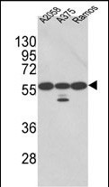
Western blot analysis of PDIA3 Antibody (Center) (Cat. #P30406) in A2058, A375, Ramos cell line lysates (35ug/lane). PDIA3 (arrow) was detected using the purified Pab.(2ug/ml)
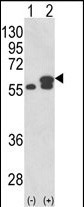
Western blot analysis of PDIA3 (arrow) using rabbit polyclonal PDIA3 Antibody (Center) (Cat. #P30406). 293 cell lysates (2 ug/lane) either nontransfected (Lane 1) or transiently transfected with the PDIA3 gene (Lane 2) .
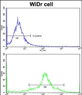
Flow cytometric analysis of widr cells using PDIA3 Antibody (Center)(bottom histogram) compared to a negative control cell (top histogram)FITC-conjugated goat-anti-rabbit secondary antibodies were used for the analysis.
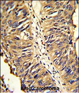
Formalin-fixed and paraffin-embedded human lung carcinoma reacted with PDIA3 Antibody (Center), which was peroxidase-conjugated to the secondary antibody, followed by DAB staining. This data demonstrates the use of this antibody for immunohistochemistry; clinical relevance has not been evaluated.
Rabbit Polyclonal Antibody to PDIA3
-
货号:
P30406 -
别名:
Protein disulfide-isomerase A3, 58 kDa glucose-regulated protein, 58 kDa microsomal protein, p58, Disulfide isomerase ER-60, Endoplasmic reticulum resident protein 57, ER protein 57, ERp57, Endoplasmic reticulum resident protein 60, ER protein 60, ERp60, PDIA3, ERP57, ERP60, GRP58 -
应用:
WB,IHC-P,FCM -
反应种属:
Human -
抗体类型:
Primary antibody -
Swissprot:
P30101 -
规格:
-
数量:
-+ -
说明书:

目录价¥1980


Rabbit Polyclonal Antibody to PDIA3
Description |
|---|
PDIA3 is the endoplasmic reticulum that interacts with lectin chaperones calreticulin and calnexin to modulate folding of newly synthesized glycoproteins. The protein was once thought to be a phospholipase; however, it has been demonstrated that the protein actually has protein disulfide isomerase activity. It is thought that complexes of lectins and this protein mediate protein folding by promoting formation of disulfide bonds in their glycoprotein substrates. |
Specification |
|
|---|---|
| Aliases | Protein disulfide-isomerase A3, 58 kDa glucose-regulated protein, 58 kDa microsomal protein, p58, Disulfide isomerase ER-60, Endoplasmic reticulum resident protein 57, ER protein 57, ERp57, Endoplasmic reticulum resident protein 60, ER protein 60, ERp60, PDIA3, ERP57, ERP60, GRP58 |
| Entrez GeneID | 2923 |
| Swissprot | P30101 |
| WB Predicted band size | 56.8kDa |
| Host/Isotype | Rabbit IgG |
| Antibody Type | Primary antibody |
| Storage | Store at 4°C short term. Aliquot and store at -20°C long term. Avoid freeze/thaw cycles. |
| Species Reactivity | Human |
| Immunogen | This PDIA3 antibody is generated from rabbits immunized with a KLH conjugated synthetic peptide between 192-220 amino acids from the Central region of human PDIA3. |
| Formulation | Purified antibody in PBS with 0.05% sodium azide,1%BSA and 50% glycerol.prepared by Saturated Ammonium Sulfate (SAS) . |
Application |
|
|---|---|
| WB | 1/1000 |
| IHC | 1/100-1/500 |
| FCM | 1/10-1/50 |
Product Image
- Western blot analysis of PDIA3 Antibody (Center) (Cat. #P30406) in A2058, A375, Ramos cell line lysates (35ug/lane). PDIA3 (arrow) was detected using the purified Pab.(2ug/ml)
- Western blot analysis of PDIA3 (arrow) using rabbit polyclonal PDIA3 Antibody (Center) (Cat. #P30406). 293 cell lysates (2 ug/lane) either nontransfected (Lane 1) or transiently transfected with the PDIA3 gene (Lane 2) .
- Flow cytometric analysis of widr cells using PDIA3 Antibody (Center)(bottom histogram) compared to a negative control cell (top histogram)FITC-conjugated goat-anti-rabbit secondary antibodies were used for the analysis.
- Formalin-fixed and paraffin-embedded human lung carcinoma reacted with PDIA3 Antibody (Center), which was peroxidase-conjugated to the secondary antibody, followed by DAB staining. This data demonstrates the use of this antibody for immunohistochemistry; clinical relevance has not been evaluated.
For Reseach Only
Application Key:WB - Western Blot | IHC - Immunohistochemistry | ICC - Immunocytochemistry | FCM - Flow Cytometry | ELISA - Enzyme-linked Immunosorbent Assay | IP - Immunoprecipitation
#P30406

相关产品


联系方式
CONTACT-
联系电话:
0731-88388785 -
公司邮箱:
sales@promab.cn -
公司地址:
湖南省长沙市高新开发区林语路239号顺畅产业园5楼
 官方微信
官方微信
产品中心
Product技术服务
service客户留言
message