


提醒成功


 微信/QQ登录
搜索


 首页
首页
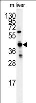
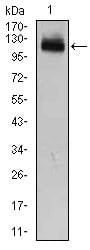
Western blot analysis of GCAT antibody (Center) (Cat.#P30356) in mouse liver tissue lysates (35ug/lane). GCAT (arrow) was detected using the purified Pab.
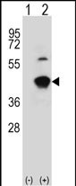
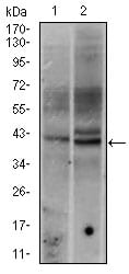
Western blot analysis of GCAT (arrow) using rabbit polyclonal GCAT Antibody (Center) (Cat.#P30356). 293 cell lysates (2 ug/lane) either nontransfected (Lane 1) or transiently transfected (Lane 2) with the GCAT gene.
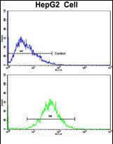
Flow cytometric analysis of HepG2 cells using GCAT Antibody (Center)(bottom histogram) compared to a negative control cell (top histogram). FITC-conjugated goat-anti-rabbit secondary antibodies were used for the analysis.
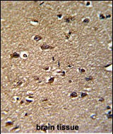
Formalin-fixed and paraffin-embedded human brain reacted with GCAT Antibody (Center), which was peroxidase-conjugated to the secondary antibody, followed by DAB staining. This data demonstrates the use of this antibody for immunohistochemistry; clinical relevance has not been evaluated.
Rabbit Polyclonal Antibody to GCAT
-
货号:
P30356 -
别名:
2-amino-3-ketobutyrate coenzyme A ligase, mitochondrial, AKB ligase, Aminoacetone synthase, Glycine acetyltransferase, GCAT, KBL -
应用:
WB,IHC-P,FCM -
反应种属:
Human, Mouse -
抗体类型:
Primary antibody -
Swissprot:
O75600 -
规格:
-
数量:
-+ -
说明书:

目录价¥1980


Rabbit Polyclonal Antibody to GCAT
Description |
|---|
The degradation of L-threonine to glycine consists of a two-step biochemical pathway involving the enzymes L-threonine dehydrogenase and 2-amino-3-ketobutyrate coenzyme A ligase. L-Threonine is first converted into 2-amino-3-ketobutyrate by L-threonine dehydrogenase. GCAT is the second enzyme in this pathway, which then catalyzes the reaction between 2-amino-3-ketobutyrate and coenzyme A to form glycine and acetyl-CoA. The enzyme is considered a class II pyridoxal-phosphate-dependent aminotransferase. |
Specification |
|
|---|---|
| Aliases | 2-amino-3-ketobutyrate coenzyme A ligase, mitochondrial, AKB ligase, Aminoacetone synthase, Glycine acetyltransferase, GCAT, KBL |
| Entrez GeneID | 23464 |
| Swissprot | O75600 |
| WB Predicted band size | 45.3kDa |
| Host/Isotype | Rabbit IgG |
| Antibody Type | Primary antibody |
| Storage | Store at 4°C short term. Aliquot and store at -20°C long term. Avoid freeze/thaw cycles. |
| Species Reactivity | Human, Mouse |
| Immunogen | This GCAT antibody is generated from rabbits immunized with a KLH conjugated synthetic peptide between 155-181 amino acids from the Central region of human GCAT. |
| Formulation | Purified antibody in PBS with 0.05% sodium azide,1%BSA and 50% glycerol.prepared by Saturated Ammonium Sulfate (SAS) . |
Application |
|
|---|---|
| WB | 1/1000 |
| IHC | 1/100-1/500 |
| FCM | 1/10-1/50 |
Product Image
- Western blot analysis of GCAT antibody (Center) (Cat.#P30356) in mouse liver tissue lysates (35ug/lane). GCAT (arrow) was detected using the purified Pab.
- Western blot analysis of GCAT (arrow) using rabbit polyclonal GCAT Antibody (Center) (Cat.#P30356). 293 cell lysates (2 ug/lane) either nontransfected (Lane 1) or transiently transfected (Lane 2) with the GCAT gene.
- Flow cytometric analysis of HepG2 cells using GCAT Antibody (Center)(bottom histogram) compared to a negative control cell (top histogram). FITC-conjugated goat-anti-rabbit secondary antibodies were used for the analysis.
- Formalin-fixed and paraffin-embedded human brain reacted with GCAT Antibody (Center), which was peroxidase-conjugated to the secondary antibody, followed by DAB staining. This data demonstrates the use of this antibody for immunohistochemistry; clinical relevance has not been evaluated.
For Reseach Only
Application Key:WB - Western Blot | IHC - Immunohistochemistry | ICC - Immunocytochemistry | FCM - Flow Cytometry | ELISA - Enzyme-linked Immunosorbent Assay | IP - Immunoprecipitation
#P30356

相关产品


联系方式
CONTACT-
联系电话:
0731-88388785 -
公司邮箱:
sales@promab.cn -
公司地址:
湖南省长沙市高新开发区林语路239号顺畅产业园5楼
 官方微信
官方微信
产品中心
Product技术服务
service客户留言
message