


提醒成功


 微信/QQ登录
搜索


 首页
首页
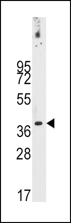
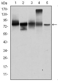
Western blot analysis of anti-PDX1 Antibody (T11) (Cat.#P30266) in NCI-H460 cell line lysates (35ug/lane).PDX1-T11(arrow) was detected using the purified Pab.
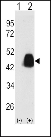
Western blot analysis of PDX1 (arrow) using PDX1 Antibody (T11) (Cat.#P30266). 293 cell lysates (2 ug/lane) either nontransfected (Lane 1) or transiently transfected with the PDX1 gene (Lane 2) (Origene Technologies).
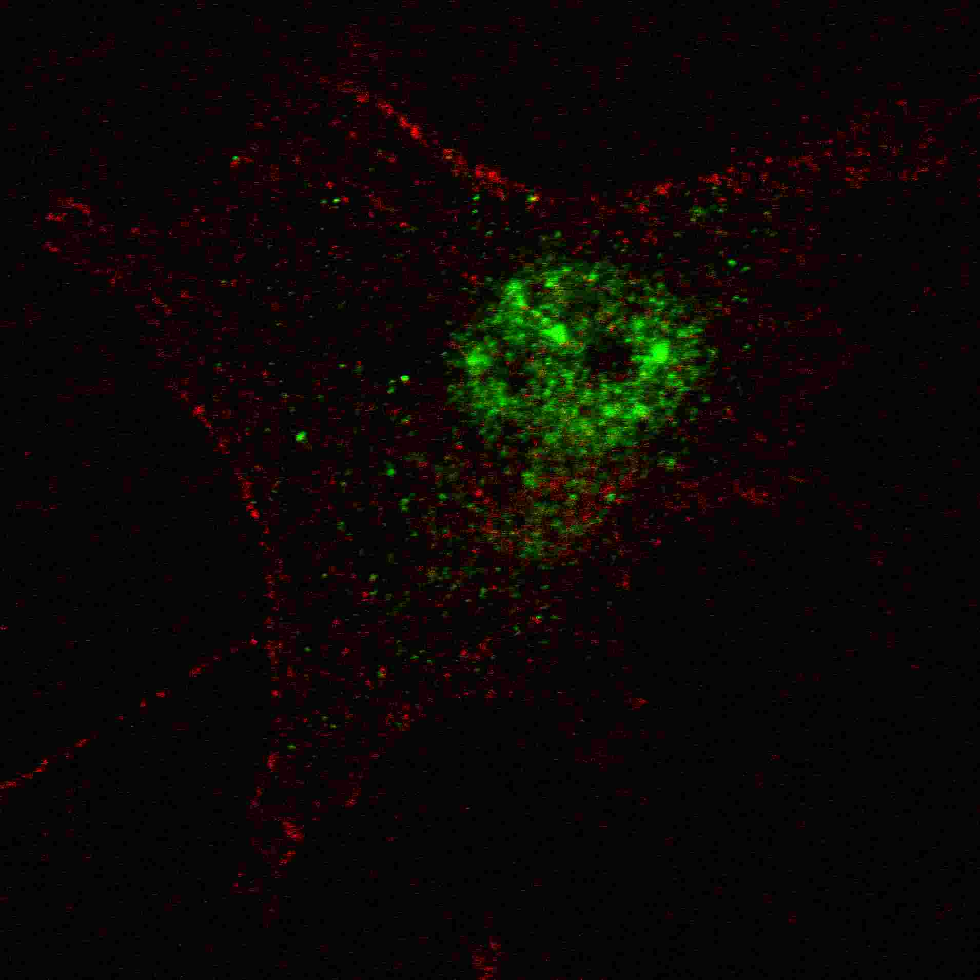
Fluorescent confocal image of SY5Y cells stained with PDX1 (T11) antibody. SY5Y cells were fixed with 4% PFA (20 min), permeabilized with Triton X-100 (0.2%, 30 min). Cells were then incubated with P30266 PDX1 (T11) primary antibody (1:100, 2 h at room temperature). For secondary antibody, Alexa Fluor? 488 conjugated donkey anti-rabbit antibody (green) was used (1:1000, 1h). Nuclei were counterstained with Hoechst 33342 (blue) (10 μg/ml, 5 min). Note the highly specific localization of the PDX1 immunosignal to the nucleus, supported by Human Protein Atlas Data (http://www.proteinatlas.org/ENSG00000110435).
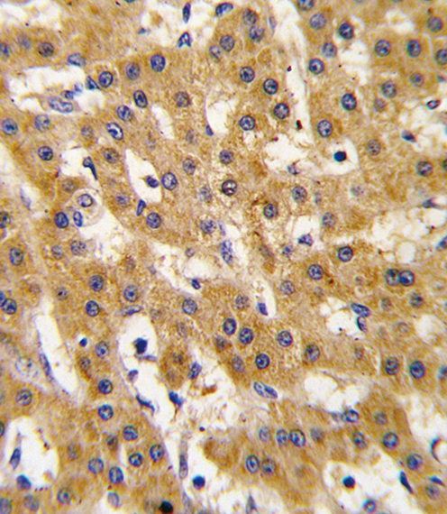
Formalin-fixed and paraffin-embedded human hepatocarcinoma tissue reacted with Phospho-PDX1-T11.ctrl antibody, which was peroxidase-conjugated to the secondary antibody, followed by DAB staining. This data demonstrates the use of this antibody for immunohistochemistry; clinical relevance has not been evaluated.
Rabbit Polyclonal Antibody to PDX1 (T11)
-
货号:
P30266 -
别名:
Pyruvate dehydrogenase protein X component, mitochondrial, Dihydrolipoamide dehydrogenase-binding protein of pyruvate dehydrogenase complex, E3-binding protein, E3BP, Lipoyl-containing pyruvate dehydrogenase complex component X, proX, PDHX, PDX1 -
应用:
WB,IHC-P,FCM -
反应种属:
Human, Mouse, Rat -
抗体类型:
Primary antibody -
Swissprot:
O00330 -
规格:
-
数量:
-+ -
说明书:

目录价¥1980


Rabbit Polyclonal Antibody to PDX1 (T11)
Description |
|---|
PDX1, located in the mitochondrial matrix, is required for anchoring dihydrolipoamide dehydrogenase (E3) to the dihydrolipoamide transacetylase (E2) core of the pyruvate dehydrogenase complexes of eukaryotes. This specific binding is essential for a functional PDH complex. Eukaryotic pyruvate dehydrogenase complexes are organized about a core consisting of the oligomeric dihydrolipoamide acetyl-transferase, around which are arranged multiple copies of pyruvate dehydrogenase, dihydrolipoamide dehydrogenase and protein X bound by noncovalent bonds. Defects in PDHX are a cause of lacticacidemia. PDX1 belongs to the 2-oxoacid dehydrogenase family and contains 1 lipoyl-binding domain. |
Specification |
|
|---|---|
| Aliases | Pyruvate dehydrogenase protein X component, mitochondrial, Dihydrolipoamide dehydrogenase-binding protein of pyruvate dehydrogenase complex, E3-binding protein, E3BP, Lipoyl-containing pyruvate dehydrogenase complex component X, proX, PDHX, PDX1 |
| Entrez GeneID | 8050 |
| Swissprot | O00330 |
| WB Predicted band size | 54.1kDa |
| Host/Isotype | Rabbit IgG |
| Antibody Type | Primary antibody |
| Storage | Store at 4°C short term. Aliquot and store at -20°C long term. Avoid freeze/thaw cycles. |
| Species Reactivity | Human, Mouse, Rat |
| Immunogen | This PDX1 antibody is generated from rabbits immunized with a KLH conjugated synthetic peptide between 1-30 amino acids from human PDX1. |
| Formulation | Purified antibody in PBS with 0.05% sodium azide. |
Application |
|
|---|---|
| WB | 1/1000 |
| IHC | 1/100-1/500 |
| FCM | 1/100 |
Product Image
- Western blot analysis of anti-PDX1 Antibody (T11) (Cat.#P30266) in NCI-H460 cell line lysates (35ug/lane).PDX1-T11(arrow) was detected using the purified Pab.
- Western blot analysis of PDX1 (arrow) using PDX1 Antibody (T11) (Cat.#P30266). 293 cell lysates (2 ug/lane) either nontransfected (Lane 1) or transiently transfected with the PDX1 gene (Lane 2) (Origene Technologies).
- Fluorescent confocal image of SY5Y cells stained with PDX1 (T11) antibody. SY5Y cells were fixed with 4% PFA (20 min), permeabilized with Triton X-100 (0.2%, 30 min). Cells were then incubated with P30266 PDX1 (T11) primary antibody (1:100, 2 h at room temperature). For secondary antibody, Alexa Fluor? 488 conjugated donkey anti-rabbit antibody (green) was used (1:1000, 1h). Nuclei were counterstained with Hoechst 33342 (blue) (10 μg/ml, 5 min). Note the highly specific localization of the PDX1 immunosignal to the nucleus, supported by Human Protein Atlas Data (http://www.proteinatlas.org/ENSG00000110435).
- Formalin-fixed and paraffin-embedded human hepatocarcinoma tissue reacted with Phospho-PDX1-T11.ctrl antibody, which was peroxidase-conjugated to the secondary antibody, followed by DAB staining. This data demonstrates the use of this antibody for immunohistochemistry; clinical relevance has not been evaluated.
For Reseach Only
Application Key:WB - Western Blot | IHC - Immunohistochemistry | ICC - Immunocytochemistry | FCM - Flow Cytometry | ELISA - Enzyme-linked Immunosorbent Assay | IP - Immunoprecipitation
#P30266

相关产品


联系方式
CONTACT-
联系电话:
0731-88388785 -
公司邮箱:
sales@promab.cn -
公司地址:
湖南省长沙市高新开发区林语路239号顺畅产业园5楼
 官方微信
官方微信
产品中心
Product技术服务
service客户留言
message