


提醒成功


 微信/QQ登录
搜索


 首页
首页
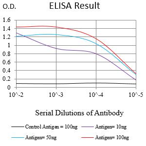
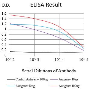
Black line: Control Antigen (100 ng);Purple line: Antigen (10ng); Blue line: Antigen (50 ng); Red line:Antigen (100 ng)
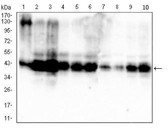
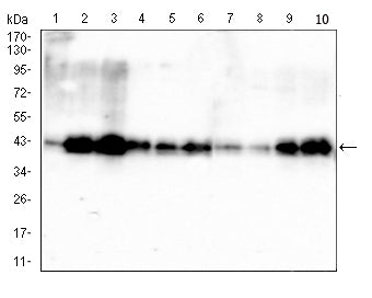
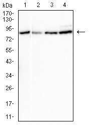
Western blot analysis using PDHA1 mouse mAb against HepG2 (1),Hek293 (2),HL-60 (3),SK-OV-3 (4),PC-3 (5),PANC-1 (6),NRK (7),C2C12 (8), C6 (9), and PC-12 (10) cell lysate.
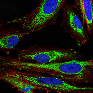
Immunofluorescence analysis of Hela cells using PDHA1 mouse mAb (green). Blue: DRAQ5 fluorescent DNA dye. Red: Actin filaments have been labeled with Alexa Fluor- 555 phalloidin. Secondary antibody from Fisher (Cat#: 35503)

Immunofluorescence analysis of NIH/3T3 cells using PDHA1 mouse mAb (green). Blue: DRAQ5 fluorescent DNA dye. Red: Actin filaments have been labeled with Alexa Fluor- 555 phalloidin. Secondary antibody from Fisher (Cat#: 35503)
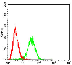
Flow cytometric analysis of Hela cells using PDHA1 mouse mAb (green) and negative control (red).
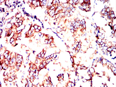
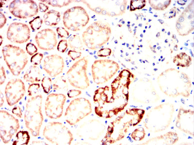
Immunohistochemical analysis of paraffin-embedded human lung cancer tissues using PDHA1 mouse mAb with DAB staining.
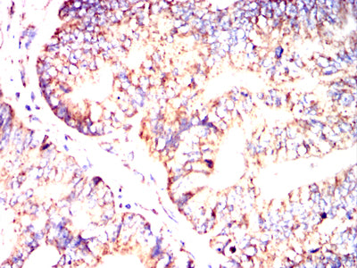
Immunohistochemical analysis of paraffin-embedded human colon cancer tissues using PDHA1 mouse mAb with DAB staining.
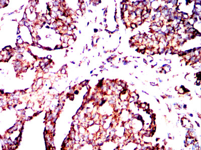
Immunohistochemical analysis of paraffin-embedded human breast cancer tissues using PDHA1 mouse mAb with DAB staining.
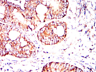
Immunohistochemical analysis of paraffin-embedded human rectum cancer tissues using PDHA1 mouse mAb with DAB staining.
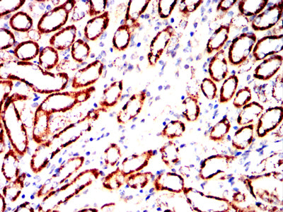
Immunohistochemical analysis of paraffin-embedded mouse kidney tissues using PDHA1 mouse mAb with DAB staining.

Immunohistochemical analysis of paraffin-embedded Rat kidney tissues using PDHA1 mouse mAb with DAB staining.
Immunohistochemical analysis of paraffin-embedded Rabbit kidney tissues using PDHA1 mouse mAb with DAB staining.
Mouse Monoclonal Antibody to PDHA1
-
货号:
32417 -
别名:
PDHA; PDHAD; PHE1A; PDHCE1A -
应用:
WB,IHC,IF,FCM -
反应种属:
Human, Mouse, Rat -
抗体类型:
Primary antibody -
Swissprot:
P08559 -
规格:
-
数量:
-+ -
说明书:

目录价¥2180


Mouse Monoclonal Antibody to PDHA1
Description |
|---|
The pyruvate dehydrogenase (PDH) complex is a nuclear-encoded mitochondrial multienzyme complex that catalyzes the overall conversion of pyruvate to acetyl-CoA and CO(2), and provides the primary link between glycolysis and the tricarboxylic acid (TCA) cycle. The PDH complex is composed of multiple copies of three enzymatic components: pyruvate dehydrogenase (E1), dihydrolipoamide acetyltransferase (E2) and lipoamide dehydrogenase (E3). The E1 enzyme is a heterotetramer of two alpha and two beta subunits. This gene encodes the E1 alpha 1 subunit containing the E1 active site, and plays a key role in the function of the PDH complex. Mutations in this gene are associated with pyruvate dehydrogenase E1-alpha deficiency and X-linked Leigh syndrome. Alternatively spliced transcript variants encoding different isoforms have been found for this gene. |
References |
|---|
| 1.Eur J Paediatr Neurol. 2021 Mar;31:27-30. 2.Pathol Res Pract. 2019 Mar;215(3):478-482. |
Specification |
|
|---|---|
| Aliases | PDHA; PDHAD; PHE1A; PDHCE1A |
| Entrez GeneID | 5160 |
| Swissprot | P08559 |
| clone | 4E12D6 |
| WB Predicted band size | 43 kDa |
| Host/Isotype | Mouse IgG2a |
| Antibody Type | Primary antibody |
| Storage | Store at 4°C short term. Aliquot and store at -20°C long term. Avoid freeze/thaw cycles. |
| Species Reactivity | Human, Mouse, Rat |
| Immunogen | Purified recombinant fragment of human PDHA1(AA: 241-390) expressed in E. Coli. |
| Formulation | Purified antibody in PBS with 0.05% sodium azide |
Application |
|
|---|---|
| WB | 1/500 - 1/2000 |
| IHC | 1/200 - 1/1000 |
| IF/ICC | 1/50 - 1/200 |
| FCM | 1/200 - 1/400 |
| ELISA | 1/10000 |
Product Image
- Black line: Control Antigen (100 ng);Purple line: Antigen (10ng); Blue line: Antigen (50 ng); Red line:Antigen (100 ng)
- Western blot analysis using PDHA1 mouse mAb against HepG2 (1),Hek293 (2),HL-60 (3),SK-OV-3 (4),PC-3 (5),PANC-1 (6),NRK (7),C2C12 (8), C6 (9), and PC-12 (10) cell lysate.
- Immunofluorescence analysis of Hela cells using PDHA1 mouse mAb (green). Blue: DRAQ5 fluorescent DNA dye. Red: Actin filaments have been labeled with Alexa Fluor- 555 phalloidin. Secondary antibody from Fisher (Cat#: 35503)
- Immunofluorescence analysis of NIH/3T3 cells using PDHA1 mouse mAb (green). Blue: DRAQ5 fluorescent DNA dye. Red: Actin filaments have been labeled with Alexa Fluor- 555 phalloidin. Secondary antibody from Fisher (Cat#: 35503)
- Flow cytometric analysis of Hela cells using PDHA1 mouse mAb (green) and negative control (red).
- Immunohistochemical analysis of paraffin-embedded human lung cancer tissues using PDHA1 mouse mAb with DAB staining.
- Immunohistochemical analysis of paraffin-embedded human colon cancer tissues using PDHA1 mouse mAb with DAB staining.
- Immunohistochemical analysis of paraffin-embedded human breast cancer tissues using PDHA1 mouse mAb with DAB staining.
- Immunohistochemical analysis of paraffin-embedded human rectum cancer tissues using PDHA1 mouse mAb with DAB staining.
- Immunohistochemical analysis of paraffin-embedded mouse kidney tissues using PDHA1 mouse mAb with DAB staining.
- Immunohistochemical analysis of paraffin-embedded Rat kidney tissues using PDHA1 mouse mAb with DAB staining.
- Immunohistochemical analysis of paraffin-embedded Rabbit kidney tissues using PDHA1 mouse mAb with DAB staining.
For Reseach Only
Application Key:WB - Western Blot | IHC - Immunohistochemistry | ICC - Immunocytochemistry | FCM - Flow Cytometry | ELISA - Enzyme-linked Immunosorbent Assay | IP - Immunoprecipitation
#32417

相关产品


联系方式
CONTACT-
联系电话:
0731-88388785 -
公司邮箱:
sales@promab.cn -
公司地址:
湖南省长沙市高新开发区林语路239号顺畅产业园5楼
 官方微信
官方微信
产品中心
Product技术服务
service客户留言
message