


提醒成功


 微信/QQ登录
搜索


 首页
首页
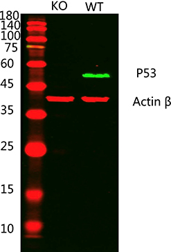
Western blot analysis of lysates from 1)p53 knockout A431 cell , 2)A431 cells, (Green) primary antibody was diluted at 1:1000, 4°Cover night, Dylight 800 secondary antibody was diluted at 1:10000, 37°C 1hour. (Red) Actin β Monoclonal Antibody(5B7) antibody was diluted at 1:5000 as loading control, 4°C over night, Dylight 680 secondary antibody was diluted at 1:10000, 37°C 1hour.

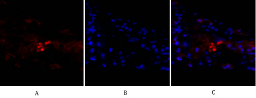
Immunofluorescence analysis of human-liver tissue. 1,p53 Polyclonal Antibody(red) was diluted at 1:200(4°C,overnight). 2, Cy3 labled Secondary antibody was diluted at 1:300(room temperature, 50min).3, Picture B: DAPI(blue) 10min. Picture A:Target. Picture B: DAPI. Picture C: merge of A+B
Immunofluorescence analysis of human-lung tissue. 1,p53 Polyclonal Antibody(red) was diluted at 1:200(4°C,overnight). 2, Cy3 labled Secondary antibody was diluted at 1:300(room temperature, 50min).3, Picture B: DAPI(blue) 10min. Picture A:Target. Picture B: DAPI. Picture C: merge of A+B
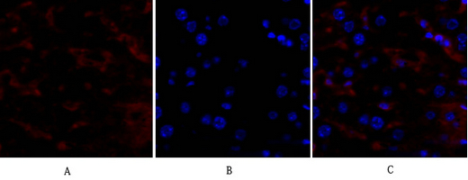
Immunofluorescence analysis of mouse-liver tissue. 1,p53 Polyclonal Antibody(red) was diluted at 1:200(4°C,overnight). 2, Cy3 labled Secondary antibody was diluted at 1:300(room temperature, 50min).3, Picture B: DAPI(blue) 10min. Picture A:Target. Picture B: DAPI. Picture C: merge of A+B
Immunohistochemical analysis of paraffin-embedded Human-lung-cancer tissue. 1,p53 Polyclonal Antibody was diluted at 1:200(4°C,overnight). 2, Sodium citrate pH 6.0 was used for antibody retrieval(>98°C,20min). 3,Secondary antibody was diluted at 1:200(room tempeRature, 30min). Negative control was used by secondary antibody only.
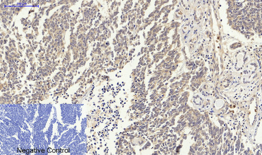
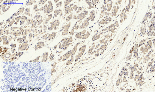
Immunohistochemical analysis of paraffin-embedded Human-stomach-cancer tissue. 1,p53 Polyclonal Antibody was diluted at 1:200(4°C,overnight). 2, Sodium citrate pH 6.0 was used for antibody retrieval(>98°C,20min). 3,Secondary antibody was diluted at 1:200(room tempeRature, 30min). Negative control was used by secondary antibody only.
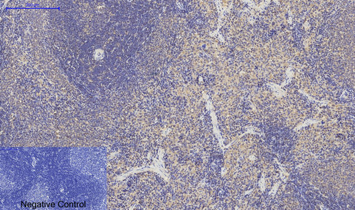
Immunohistochemical analysis of paraffin-embedded Rat-spleen tissue. 1,p53 Polyclonal Antibody was diluted at 1:200(4°C,overnight). 2, Sodium citrate pH 6.0 was used for antibody retrieval(>98°C,20min). 3,Secondary antibody was diluted at 1:200(room tempeRature, 30min). Negative control was used by secondary antibody only.
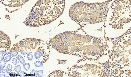
Immunohistochemical analysis of paraffin-embedded Mouse-testis tissue. 1,p53 Polyclonal Antibody was diluted at 1:200(4°C,overnight). 2, Sodium citrate pH 6.0 was used for antibody retrieval(>98°C,20min). 3,Secondary antibody was diluted at 1:200(room tempeRature, 30min). Negative control was used by secondary antibody only.
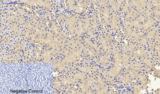
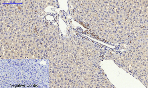
Immunohistochemical analysis of paraffin-embedded Mouse-liver tissue. 1,p53 Polyclonal Antibody was diluted at 1:200(4°C,overnight). 2, Sodium citrate pH 6.0 was used for antibody retrieval(>98°C,20min). 3,Secondary antibody was diluted at 1:200(room tempeRature, 30min). Negative control was used by secondary antibody only.
Immunohistochemical analysis of paraffin-embedded Mouse-kidney tissue. 1,p53 Polyclonal Antibody was diluted at 1:200(4°C,overnight). 2, Sodium citrate pH 6.0 was used for antibody retrieval(>98°C,20min). 3,Secondary antibody was diluted at 1:200(room tempeRature, 30min). Negative control was used by secondary antibody only.
Rabbit Polyclonal Antibody to p53(Ab-15)
-
货号:
P41312 -
别名:
Tumor suppressor p53; Phosphoprotein p53; Antigen NY-CO-13; TP53; -
应用:
WB,IHC,IF -
反应种属:
Human,Mouse,Rat -
抗体类型:
Primary antibody -
Swissprot:
P04637 -
规格:
-
数量:
-+ -
说明书:

目录价¥1980


Rabbit Polyclonal Antibody to p53(Ab-15)
Description |
|---|
p53 is a nuclear protein which plays an essential role in the regulation of cell cycle specifically in the transition from G0 to G1. It is found in very low levels in normal cells however in a variety of transformed cell lines in high amounts and believed to contribute to transformation and malignancy. The open reading frame of p53 is 393 amino acids long, with the central region (consisting of amino acids from about 100 to 300) containing the DNA-binding domain. This proteolysis-resistant core is flanked by a C-terminal end mediating oligomerization and an N-terminal end containing a strong transcription activation signal. p53 binds as a tetramer to a PBS (p53-Binding Site) and activates the expression of downstream genes that inhibit growth and/or invasion. p53 binds as a tetramer to a p53-binding site (PBS) and to activate the expression of adjacent genes that inhibit growth and/or invasion. Deletion of one or both p53 alleles reduces the expression of tetramers, resulting in decreased expression of the growth inhibitory genes |
References |
|---|
| Lin T, et al. (2005) Nat Cell Biol; 7(2): 165-71. Vega FM, et al. (2004) Mol Cell Biol; 24(23): 10366-80. Li J, et al. (2004) J Biol Chem; 279(40): 41275-9. Wang J, et al. (2004) J Biol Chem; 279(38): 39584-92. |
Specification |
|
|---|---|
| Aliases | Tumor suppressor p53; Phosphoprotein p53; Antigen NY-CO-13; TP53; |
| Entrez GeneID | 7157; |
| Swissprot | P04637 |
| Host/Isotype | Rabbit IgG |
| Antibody Type | Primary antibody |
| Storage | Store at 4°C short term. Aliquot and store at -20°C long term. Avoid freeze/thaw cycles. |
| Species Reactivity | Human,Mouse,Rat |
| Immunogen | Peptide sequence around aa.13~17 (P-L-S-Q-E) derived from Human p53. |
| Formulation | Liquid in PBS containing 50% glycerol, 0.5% BSA and 0.02% sodium azide. |
Application |
|
|---|---|
| WB | 1/500-1/2000 |
| IHC | 1/100-1/300 |
| IF/ICC | 1/200-1/1000 |
Product Image
- Western blot analysis of lysates from 1)p53 knockout A431 cell , 2)A431 cells, (Green) primary antibody was diluted at 1:1000, 4°Cover night, Dylight 800 secondary antibody was diluted at 1:10000, 37°C 1hour. (Red) Actin β Monoclonal Antibody(5B7) antibody was diluted at 1:5000 as loading control, 4°C over night, Dylight 680 secondary antibody was diluted at 1:10000, 37°C 1hour.
- Immunofluorescence analysis of human-liver tissue. 1,p53 Polyclonal Antibody(red) was diluted at 1:200(4°C,overnight). 2, Cy3 labled Secondary antibody was diluted at 1:300(room temperature, 50min).3, Picture B: DAPI(blue) 10min. Picture A:Target. Picture B: DAPI. Picture C: merge of A+B
- Immunofluorescence analysis of human-lung tissue. 1,p53 Polyclonal Antibody(red) was diluted at 1:200(4°C,overnight). 2, Cy3 labled Secondary antibody was diluted at 1:300(room temperature, 50min).3, Picture B: DAPI(blue) 10min. Picture A:Target. Picture B: DAPI. Picture C: merge of A+B
- Immunofluorescence analysis of mouse-liver tissue. 1,p53 Polyclonal Antibody(red) was diluted at 1:200(4°C,overnight). 2, Cy3 labled Secondary antibody was diluted at 1:300(room temperature, 50min).3, Picture B: DAPI(blue) 10min. Picture A:Target. Picture B: DAPI. Picture C: merge of A+B
- Immunohistochemical analysis of paraffin-embedded Human-lung-cancer tissue. 1,p53 Polyclonal Antibody was diluted at 1:200(4°C,overnight). 2, Sodium citrate pH 6.0 was used for antibody retrieval(>98°C,20min). 3,Secondary antibody was diluted at 1:200(room tempeRature, 30min). Negative control was used by secondary antibody only.
- Immunohistochemical analysis of paraffin-embedded Human-stomach-cancer tissue. 1,p53 Polyclonal Antibody was diluted at 1:200(4°C,overnight). 2, Sodium citrate pH 6.0 was used for antibody retrieval(>98°C,20min). 3,Secondary antibody was diluted at 1:200(room tempeRature, 30min). Negative control was used by secondary antibody only.
- Immunohistochemical analysis of paraffin-embedded Rat-spleen tissue. 1,p53 Polyclonal Antibody was diluted at 1:200(4°C,overnight). 2, Sodium citrate pH 6.0 was used for antibody retrieval(>98°C,20min). 3,Secondary antibody was diluted at 1:200(room tempeRature, 30min). Negative control was used by secondary antibody only.
- Immunohistochemical analysis of paraffin-embedded Mouse-testis tissue. 1,p53 Polyclonal Antibody was diluted at 1:200(4°C,overnight). 2, Sodium citrate pH 6.0 was used for antibody retrieval(>98°C,20min). 3,Secondary antibody was diluted at 1:200(room tempeRature, 30min). Negative control was used by secondary antibody only.
- Immunohistochemical analysis of paraffin-embedded Mouse-liver tissue. 1,p53 Polyclonal Antibody was diluted at 1:200(4°C,overnight). 2, Sodium citrate pH 6.0 was used for antibody retrieval(>98°C,20min). 3,Secondary antibody was diluted at 1:200(room tempeRature, 30min). Negative control was used by secondary antibody only.
- Immunohistochemical analysis of paraffin-embedded Mouse-kidney tissue. 1,p53 Polyclonal Antibody was diluted at 1:200(4°C,overnight). 2, Sodium citrate pH 6.0 was used for antibody retrieval(>98°C,20min). 3,Secondary antibody was diluted at 1:200(room tempeRature, 30min). Negative control was used by secondary antibody only.
For Reseach Only
Application Key:WB - Western Blot | IHC - Immunohistochemistry | ICC - Immunocytochemistry | FCM - Flow Cytometry | ELISA - Enzyme-linked Immunosorbent Assay | IP - Immunoprecipitation
#P41312

相关产品


联系方式
CONTACT-
联系电话:
0731-88388785 -
公司邮箱:
sales@promab.cn -
公司地址:
湖南省长沙市高新开发区林语路239号顺畅产业园5楼
 官方微信
官方微信
产品中心
Product技术服务
service客户留言
message