


提醒成功


 微信/QQ登录
搜索


 首页
首页
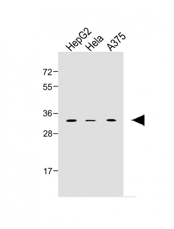
All lanes : Anti-MSX1 Antibody (Center) at 1:1000 dilution
Lane 1: HepG2 MG whole cell lysate
Lane 2: Hela whole cell lysate
Lane 3: A375 whole cell lysate
Lysates/proteins at 20 µg per lane.
Secondary
Goat Anti-Rabbit IgG, (H+L), Peroxidase conjugated at 1/10000 dilution.
Observed band size : 32kDa
Blocking/Dilution buffer: 5% NFDM/TBST.
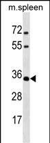
MSX1 Antibody (Center) (Cat. #P34893) western blot analysis in mouse spleen tissue lysates (35ug/lane).This demonstrates the MSX1 antibody detected the MSX1 protein (arrow).
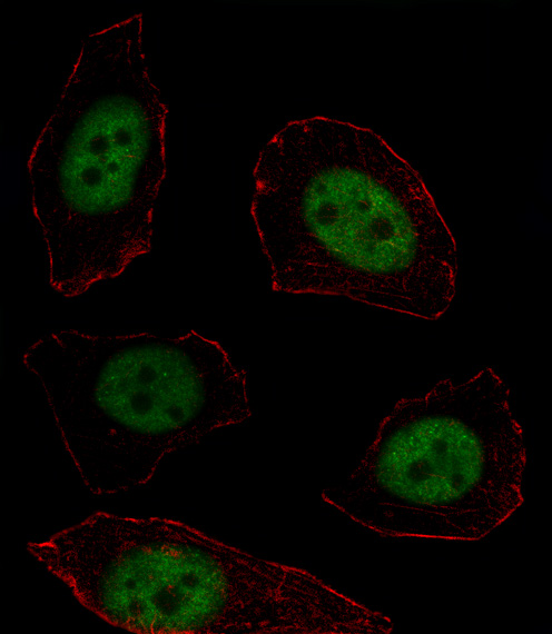
Fluorescent image of U251 cell stained with MSX1 Antibody (Center)(Cat#P34893).U251 cells were fixed with 4% PFA (20 min), permeabilized with Triton X-100 (0.1%, 10 min), then incubated with MSX1 primary antibody (1:25, 1 h at 37℃). For secondary antibody, Alexa Fluor® 488 conjugated donkey anti-rabbit antibody (green) was used (1:400, 50 min at 37℃).Cytoplasmic actin was counterstained with Alexa Fluor® 555 (red) conjugated Phalloidin (7units/ml, 1 h at 37℃).MSX1 immunoreactivity is localized to Nucleus significantly.
Rabbit Polyclonal Antibody to MSX1
-
货号:
P34893 -
别名:
Homeobox protein MSX-1, Homeobox protein Hox-7, Msh homeobox 1-like protein, MSX1, HOX7 -
应用:
WB,IF -
反应种属:
Human, Mouse, Rat -
抗体类型:
Primary antibody -
Swissprot:
P28360 -
规格:
-
数量:
-+ -
说明书:

目录价¥1980


Rabbit Polyclonal Antibody to MSX1
Description |
|---|
This gene encodes a member of the muscle segment homeobox gene family. The encoded protein functions as a transcriptional repressor during embryogenesis through interactions with components of the core transcription complex and other homeoproteins. It may also have roles in limb-pattern formation, craniofacial development, particularly odontogenesis, and tumor growth inhibition. Mutations in this gene, which was once known as homeobox 7, have been associated with nonsyndromic cleft lip with or without cleft palate 5, Witkop syndrome, Wolf-Hirschom syndrome, and autosomoal dominant hypodontia. |
Specification |
|
|---|---|
| Aliases | Homeobox protein MSX-1, Homeobox protein Hox-7, Msh homeobox 1-like protein, MSX1, HOX7 |
| Entrez GeneID | 4487 |
| Swissprot | P28360 |
| WB Predicted band size | 31.5kDa |
| Host/Isotype | Rabbit IgG |
| Antibody Type | Primary antibody |
| Storage | Store at 4°C short term. Aliquot and store at -20°C long term. Avoid freeze/thaw cycles. |
| Species Reactivity | Human, Mouse, Rat |
| Immunogen | This MSX1 antibody is generated from rabbits immunized with a KLH conjugated synthetic peptide between 111-138 amino acids from the Central region of human MSX1. |
| Formulation | Purified antibody in PBS with 0.05% sodium azide. |
Application |
|
|---|---|
| WB | 1/1000-1/2000 |
| IF/ICC | 1/10-1/50 |
Product Image
- All lanes : Anti-MSX1 Antibody (Center) at 1:1000 dilution Lane 1: HepG2 MG whole cell lysate Lane 2: Hela whole cell lysate Lane 3: A375 whole cell lysate Lysates/proteins at 20 µg per lane. Secondary Goat Anti-Rabbit IgG, (H+L), Peroxidase conjugated at 1/10000 dilution. Observed band size : 32kDa Blocking/Dilution buffer: 5% NFDM/TBST.
- MSX1 Antibody (Center) (Cat. #P34893) western blot analysis in mouse spleen tissue lysates (35ug/lane).This demonstrates the MSX1 antibody detected the MSX1 protein (arrow).
- Fluorescent image of U251 cell stained with MSX1 Antibody (Center)(Cat#P34893).U251 cells were fixed with 4% PFA (20 min), permeabilized with Triton X-100 (0.1%, 10 min), then incubated with MSX1 primary antibody (1:25, 1 h at 37℃). For secondary antibody, Alexa Fluor® 488 conjugated donkey anti-rabbit antibody (green) was used (1:400, 50 min at 37℃).Cytoplasmic actin was counterstained with Alexa Fluor® 555 (red) conjugated Phalloidin (7units/ml, 1 h at 37℃).MSX1 immunoreactivity is localized to Nucleus significantly.
For Reseach Only
Application Key:WB - Western Blot | IHC - Immunohistochemistry | ICC - Immunocytochemistry | FCM - Flow Cytometry | ELISA - Enzyme-linked Immunosorbent Assay | IP - Immunoprecipitation
#P34893

相关产品


联系方式
CONTACT-
联系电话:
0731-88388785 -
公司邮箱:
sales@promab.cn -
公司地址:
湖南省长沙市高新开发区林语路239号顺畅产业园5楼
 官方微信
官方微信
产品中心
Product技术服务
service客户留言
message