


提醒成功


 微信/QQ登录
搜索


 首页
首页
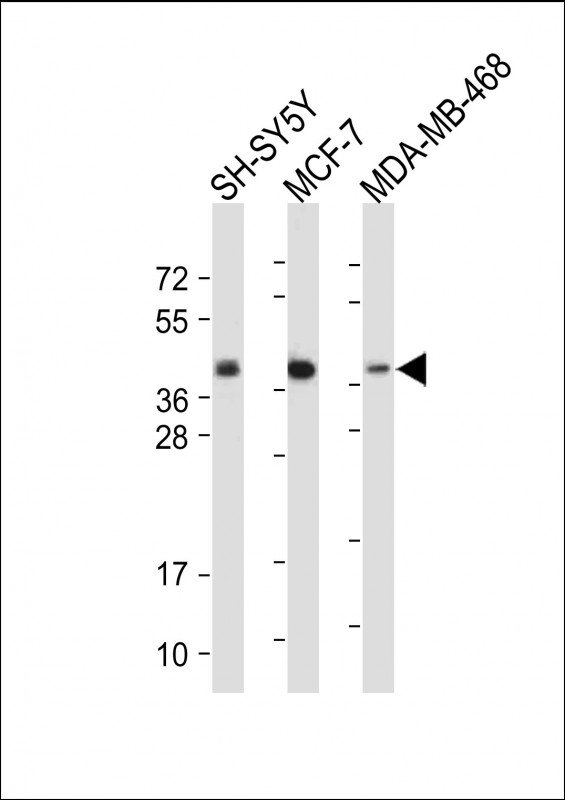
All lanes : Anti-CREB(S133) Antibody at 1:2000 dilution
Lane 1: SH-SY5Y whole cell lysate
Lane 2: MCF-7 whole cell lysate
Lane 3: MDA-MB-468 whole cell lysate
Lysates/proteins at 20 µg per lane.
Secondary
Goat Anti-Rabbit IgG, (H+L), Peroxidase conjugated at 1/10000 dilution.
Predicted band size : 37 kDa
Blocking/Dilution buffer: 5% NFDM/TBST.
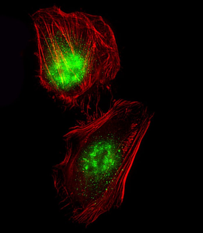
Immunofluorescent analysis of 4% paraformaldehyde-fixed, 0. 1% Triton X-100 permeabilized Hela cells labeling CREB1 with P34708 at 1/25 dilution, followed by Dylight® 488-conjugated goat anti-Rabbit IgG secondary antibody at 1/200 dilution (green). Immunofluorescence image showing Nucleus and Weak Cytoplasm staining on Hela cell line. Cytoplasmic actin is detected with Dylight® 554 Phalloidin(red). The nuclear counter stain is DAPI (blue).
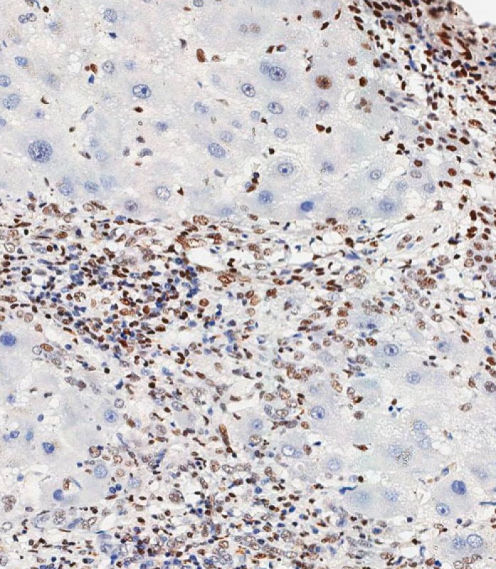
Immunohistochemical analysis of paraffin-embedded Human hepatocarcinoma tissue using P34708 performed on the Leica® BOND RXm. Tissue was fixed with formaldehyde at room temperature, antigen retrieval was by heat mediation with a EDTA buffer (pH9. 0). Samples were incubated with primary antibody(1:500) for 1 hours at room temperature. A undiluted biotinylated CRF Anti-Polyvalent HRP Polymer antibody was used as the secondary antibody.
Rabbit Polyclonal Antibody to CREB(S133)
-
货号:
P34708 -
别名:
Cyclic AMP-responsive element-binding protein 1, CREB-1, cAMP-responsive element-binding protein 1, CREB1 -
应用:
WB,IHC-P,IF -
反应种属:
Human -
抗体类型:
Primary antibody -
Swissprot:
P16220 -
规格:
-
数量:
-+ -
说明书:

目录价¥1980


Rabbit Polyclonal Antibody to CREB(S133)
Description |
|---|
Phosphorylation-dependent transcription factor that stimulates transcription upon binding to the DNA cAMP response element (CRE), a sequence present in many viral and cellular promoters. Transcription activation is enhanced by the TORC coactivators which act independently of Ser-133 phosphorylation. Involved in different cellular processes including the synchronization of circadian rhythmicity and the differentiation of adipose cells. |
Specification |
|
|---|---|
| Aliases | Cyclic AMP-responsive element-binding protein 1, CREB-1, cAMP-responsive element-binding protein 1, CREB1 |
| Entrez GeneID | 1385 |
| Swissprot | P16220 |
| WB Predicted band size | 35.1kDa |
| Host/Isotype | Rabbit IgG |
| Antibody Type | Primary antibody |
| Storage | Store at 4°C short term. Aliquot and store at -20°C long term. Avoid freeze/thaw cycles. |
| Species Reactivity | Human |
| Immunogen | This CREB(S133) antibody is generated from a rabbit immunized with a KLH conjugated synthetic peptide between 110-140 from the human region of human CREB(S133). |
Application |
|
|---|---|
| WB | 1/2000 |
| IHC | 1/500 |
| IF/ICC | 1/25 |
Product Image
- All lanes : Anti-CREB(S133) Antibody at 1:2000 dilution Lane 1: SH-SY5Y whole cell lysate Lane 2: MCF-7 whole cell lysate Lane 3: MDA-MB-468 whole cell lysate Lysates/proteins at 20 µg per lane. Secondary Goat Anti-Rabbit IgG, (H+L), Peroxidase conjugated at 1/10000 dilution. Predicted band size : 37 kDa Blocking/Dilution buffer: 5% NFDM/TBST.
- Immunofluorescent analysis of 4% paraformaldehyde-fixed, 0. 1% Triton X-100 permeabilized Hela cells labeling CREB1 with P34708 at 1/25 dilution, followed by Dylight® 488-conjugated goat anti-Rabbit IgG secondary antibody at 1/200 dilution (green). Immunofluorescence image showing Nucleus and Weak Cytoplasm staining on Hela cell line. Cytoplasmic actin is detected with Dylight® 554 Phalloidin(red). The nuclear counter stain is DAPI (blue).
- Immunohistochemical analysis of paraffin-embedded Human hepatocarcinoma tissue using P34708 performed on the Leica® BOND RXm. Tissue was fixed with formaldehyde at room temperature, antigen retrieval was by heat mediation with a EDTA buffer (pH9. 0). Samples were incubated with primary antibody(1:500) for 1 hours at room temperature. A undiluted biotinylated CRF Anti-Polyvalent HRP Polymer antibody was used as the secondary antibody.
For Reseach Only
Application Key:WB - Western Blot | IHC - Immunohistochemistry | ICC - Immunocytochemistry | FCM - Flow Cytometry | ELISA - Enzyme-linked Immunosorbent Assay | IP - Immunoprecipitation
#P34708

相关产品


联系方式
CONTACT-
联系电话:
0731-88388785 -
公司邮箱:
sales@promab.cn -
公司地址:
湖南省长沙市高新开发区林语路239号顺畅产业园5楼
 官方微信
官方微信
产品中心
Product技术服务
service客户留言
message