


提醒成功


 微信/QQ登录
搜索


 首页
首页
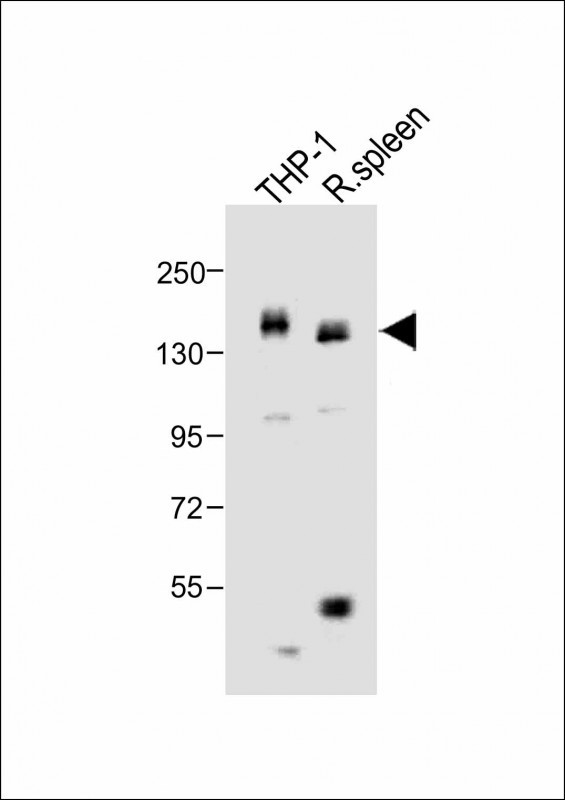
All lanes : Anti-MCSF Receptor (CSF1R) Antibody (C-term) at 1:1000 dilution
Lane 1: THP-1 whole cell lysate
Lane 2: rat spleen lysate
Lysates/proteins at 20 µg per lane.
Secondary
Goat Anti-Rabbit IgG, (H+L), Peroxidase conjugated at 1/10000 dilution.
Predicted band size : 108 kDa
Blocking/Dilution buffer: 5% NFDM/TBST.
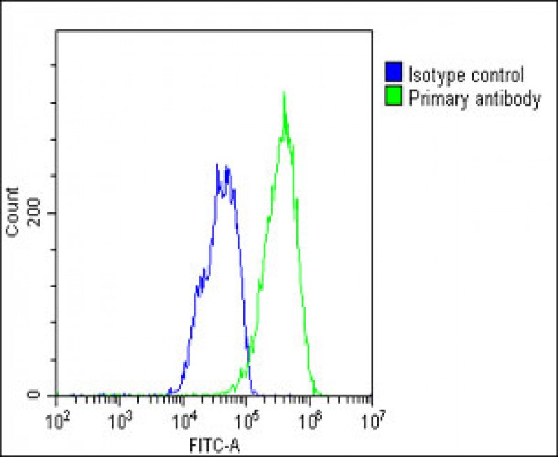
Overlay histogram showing HepG2 cells stained with P34603(green line). The cells were fixed with 2% paraformaldehyde (10 min) and then permeabilized with 90% methanol for 10 min. The cells were then icubated in 2% bovine serum albumin to block non-specific protein-protein interactions followed by the antibody (P34603, 1:25 dilution) for 60 min at 37ºC. The secondary antibody used was Goat-Anti-Rabbit IgG, DyLight® 488 Conjugated Highly Cross-Adsorbed(1583138) at 1/200 dilution for 40 min at 37ºC. Isotype control antibody (blue line) was rabbit IgG1 (1μg/1x10^6 cells) used under the same conditions. Acquisition of >10, 000 events was performed.
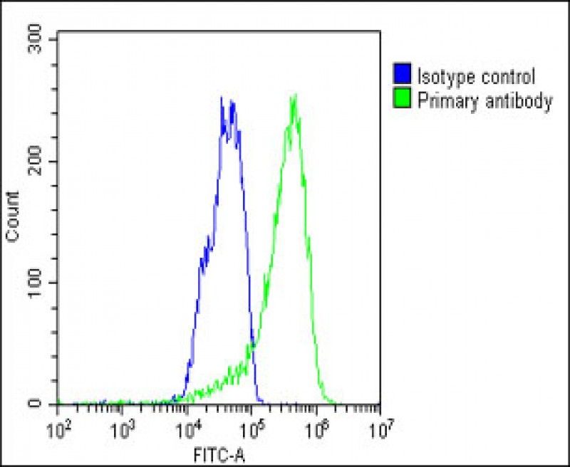
Overlay histogram showing HepG2 cells stained with P34603(green line). The cells were fixed with 2% paraformaldehyde (10 min) and then permeabilized with 90% methanol for 10 min. The cells were then icubated in 2% bovine serum albumin to block non-specific protein-protein interactions followed by the antibody (P34603, 1:25 dilution) for 60 min at 37ºC. The secondary antibody used was Goat-Anti-Rabbit IgG, DyLight® 488 Conjugated Highly Cross-Adsorbed(1583138) at 1/200 dilution for 40 min at 37ºC. Isotype control antibody (blue line) was rabbit IgG1 (1μg/1x10^6 cells) used under the same conditions. Acquisition of >10, 000 events was performed.
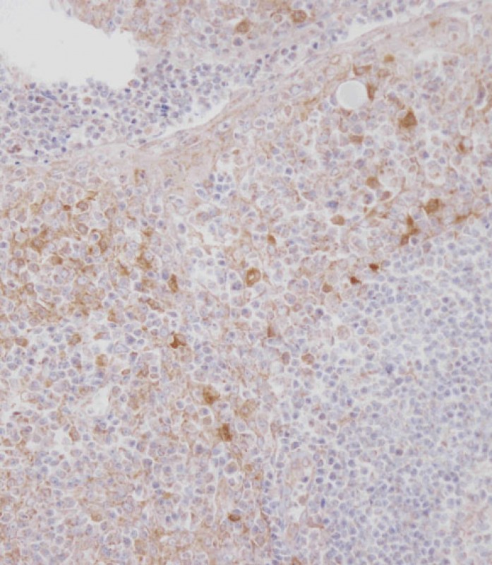
Immunohistochemical analysis of P34603 on paraffin-embedded Human tonsil tissue. Tissue was fixed with formaldehyde at room temperature. Heat induced epitope retrieval was performed by EDTA buffer (pH9. 0). Samples were incubated with primary antibody(1:100) for 1 hour at room temperature. Undiluted CRF Anti-Polyvalent HRP Polymer antibody was used as the secondary antibody.
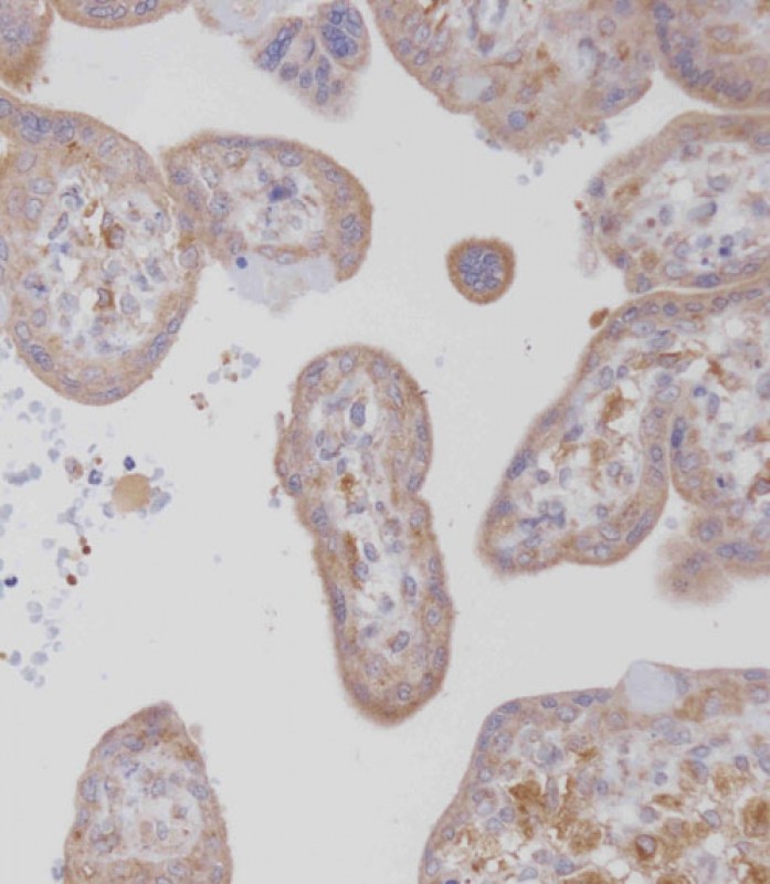
Immunohistochemical analysis of P34603 on paraffin-embedded Human placenta tissue. Tissue was fixed with formaldehyde at room temperature. Heat induced epitope retrieval was performed by EDTA buffer (pH9. 0). Samples were incubated with primary antibody(1:100) for 1 hour at room temperature. Undiluted CRF Anti-Polyvalent HRP Polymer antibody was used as the secondary antibody.
Rabbit Polyclonal Antibody to MCSF Receptor (CSF1R)
-
货号:
P34603 -
别名:
Macrophage colony-stimulating factor 1 receptor, CSF-1 receptor, CSF-1-R, CSF-1R, M-CSF-R, Proto-oncogene c-Fms, CD115, CSF1R, FMS -
应用:
WB,IHC-P,FCM -
反应种属:
Human, Mouse, Rat -
抗体类型:
Primary antibody -
Swissprot:
P07333 -
规格:
-
数量:
-+ -
说明书:

目录价¥1980


Rabbit Polyclonal Antibody to MCSF Receptor (CSF1R)
Description |
|---|
CSF1R is the receptor for colony stimulating factor 1, a cytokine which controls the production, differentiation, and function of macrophages. This receptor mediates most if not all of the biological effects of this cytokine. Ligand binding activates the receptor kinase through a process of oligomerization and transphosphorylation. This protein is a tyrosine kinase transmembrane receptor and member of the CSF1/PDGF receptor family of tyrosine-protein kinases. Mutations in the gene encoding CSF1R have been associated with a predisposition to myeloid malignancy. |
Specification |
|
|---|---|
| Aliases | Macrophage colony-stimulating factor 1 receptor, CSF-1 receptor, CSF-1-R, CSF-1R, M-CSF-R, Proto-oncogene c-Fms, CD115, CSF1R, FMS |
| Entrez GeneID | 1436 |
| Swissprot | P07333 |
| WB Predicted band size | 108.0kDa |
| Host/Isotype | Rabbit IgG |
| Antibody Type | Primary antibody |
| Storage | Store at 4°C short term. Aliquot and store at -20°C long term. Avoid freeze/thaw cycles. |
| Species Reactivity | Human, Mouse, Rat |
| Immunogen | This MCSF Receptor (CSF1R) antibody is generated from rabbits immunized with a KLH conjugated synthetic peptide between 940-971 amino acids from the C-terminal region of human MCSF Receptor (CSF1R). |
| Formulation | Purified antibody in PBS with 0.05% sodium azide,1%BSA and 50% glycerol.prepared by Saturated Ammonium Sulfate (SAS) . |
Application |
|
|---|---|
| WB | 1/1000-1/2000 |
| IHC | 1/100 |
| FCM | 1/25 |
Product Image
- All lanes : Anti-MCSF Receptor (CSF1R) Antibody (C-term) at 1:1000 dilution Lane 1: THP-1 whole cell lysate Lane 2: rat spleen lysate Lysates/proteins at 20 µg per lane. Secondary Goat Anti-Rabbit IgG, (H+L), Peroxidase conjugated at 1/10000 dilution. Predicted band size : 108 kDa Blocking/Dilution buffer: 5% NFDM/TBST.
- Overlay histogram showing HepG2 cells stained with P34603(green line). The cells were fixed with 2% paraformaldehyde (10 min) and then permeabilized with 90% methanol for 10 min. The cells were then icubated in 2% bovine serum albumin to block non-specific protein-protein interactions followed by the antibody (P34603, 1:25 dilution) for 60 min at 37ºC. The secondary antibody used was Goat-Anti-Rabbit IgG, DyLight® 488 Conjugated Highly Cross-Adsorbed(1583138) at 1/200 dilution for 40 min at 37ºC. Isotype control antibody (blue line) was rabbit IgG1 (1μg/1x10^6 cells) used under the same conditions. Acquisition of >10, 000 events was performed.
- Overlay histogram showing HepG2 cells stained with P34603(green line). The cells were fixed with 2% paraformaldehyde (10 min) and then permeabilized with 90% methanol for 10 min. The cells were then icubated in 2% bovine serum albumin to block non-specific protein-protein interactions followed by the antibody (P34603, 1:25 dilution) for 60 min at 37ºC. The secondary antibody used was Goat-Anti-Rabbit IgG, DyLight® 488 Conjugated Highly Cross-Adsorbed(1583138) at 1/200 dilution for 40 min at 37ºC. Isotype control antibody (blue line) was rabbit IgG1 (1μg/1x10^6 cells) used under the same conditions. Acquisition of >10, 000 events was performed.
- Immunohistochemical analysis of P34603 on paraffin-embedded Human tonsil tissue. Tissue was fixed with formaldehyde at room temperature. Heat induced epitope retrieval was performed by EDTA buffer (pH9. 0). Samples were incubated with primary antibody(1:100) for 1 hour at room temperature. Undiluted CRF Anti-Polyvalent HRP Polymer antibody was used as the secondary antibody.
- Immunohistochemical analysis of P34603 on paraffin-embedded Human placenta tissue. Tissue was fixed with formaldehyde at room temperature. Heat induced epitope retrieval was performed by EDTA buffer (pH9. 0). Samples were incubated with primary antibody(1:100) for 1 hour at room temperature. Undiluted CRF Anti-Polyvalent HRP Polymer antibody was used as the secondary antibody.
For Reseach Only
Application Key:WB - Western Blot | IHC - Immunohistochemistry | ICC - Immunocytochemistry | FCM - Flow Cytometry | ELISA - Enzyme-linked Immunosorbent Assay | IP - Immunoprecipitation
#P34603

相关产品


联系方式
CONTACT-
联系电话:
0731-88388785 -
公司邮箱:
sales@promab.cn -
公司地址:
湖南省长沙市高新开发区林语路239号顺畅产业园5楼
 官方微信
官方微信
产品中心
Product技术服务
service客户留言
message