


提醒成功


 微信/QQ登录
搜索


 首页
首页
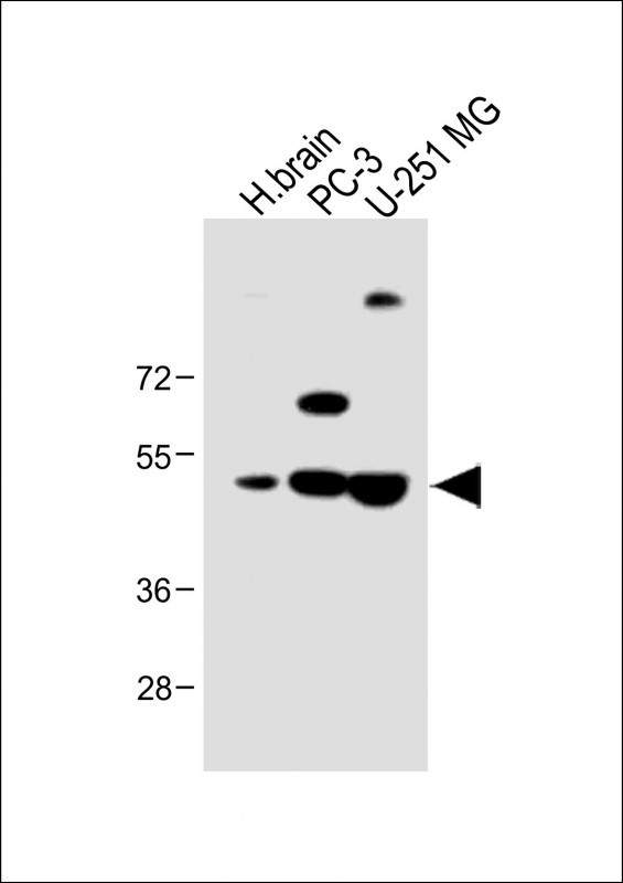
All lanes : Anti-AP1M1 Antibody (Center) at 1:1000 dilution
Lane 1: Human brain lysate
Lane 2: PC-3 whole cell lysate
Lane 3: U-251 MG whole cell lysate
Lysates/proteins at 20 µg per lane.
Secondary
Goat Anti-Rabbit IgG, (H+L), Peroxidase conjugated at 1/10000 dilution.
Predicted band size : 49 kDa
Blocking/Dilution buffer: 5% NFDM/TBST.

Immunofluorescent analysis of 4% paraformaldehyde-fixed, 0. 1% Triton X-100 permeabilized U-251 MG cells labeling AP1M1 with P34586 at 1/25 dilution, followed by Dylight® 488-conjugated goat anti-Rabbit IgG secondary antibody at 1/200 dilution (green). Immunofluorescence image showing Cytoplasm and Weak Nucleus staining on U-251 MG cell line. Cytoplasmic actin is detected with Dylight® 554 Phalloidin(red). The nuclear counter stain is DAPI (blue).

Overlay histogram showing U-251 MG cells stained with P34586(green line). The cells were fixed with 2% paraformaldehyde (10 min) and then permeabilized with 90% methanol for 10 min. The cells were then icubated in 2% bovine serum albumin to block non-specific protein-protein interactions followed by the antibody (P34586, 1:25 dilution) for 60 min at 37ºC. The secondary antibody used was Goat-Anti-Rabbit IgG, DyLight® 488 Conjugated Highly Cross-Adsorbed(1583138) at 1/200 dilution for 40 min at 37ºC. Isotype control antibody (blue line) was rabbit IgG1 (1μg/1x10^6 cells) used under the same conditions. Acquisition of >10, 000 events was performed.
Rabbit Polyclonal Antibody to AP1M1
-
货号:
P34586 -
别名:
AP-1 complex subunit mu-1, AP-mu chain family member mu1A, Adaptor protein complex AP-1 subunit mu-1, Adaptor-related protein complex 1 subunit mu-1, Clathrin assembly protein complex 1 mu-1 medium chain 1, Clathrin coat assembly protein AP47, Clathrin coat-associated protein AP47, Golgi adaptor HA1/AP1 adaptin mu-1 subunit, Mu-adaptin 1, Mu1A-adaptin, AP1M1, CLTNM -
应用:
WB,IF,FCM -
反应种属:
Human, Mouse, Rat -
抗体类型:
Primary antibody -
Swissprot:
Q9BXS5 -
规格:
-
数量:
-+ -
说明书:

目录价¥1980


Rabbit Polyclonal Antibody to AP1M1
Description |
|---|
The protein encoded by this gene is the medium chain of the trans-Golgi network clathrin-associated protein complex AP-1. The other components of this complex are beta-prime-adaptin, gamma-adaptin, and the small chain AP1S1. This complex is located at the Golgi vesicle and links clathrin to receptors in coated vesicles. These vesicles are involved in endocytosis and Golgi processing. Alternatively spliced transcript variants encoding distinct protein isoforms have been found for this gene. [provided by RefSeq]. |
Specification |
|
|---|---|
| Aliases | AP-1 complex subunit mu-1, AP-mu chain family member mu1A, Adaptor protein complex AP-1 subunit mu-1, Adaptor-related protein complex 1 subunit mu-1, Clathrin assembly protein complex 1 mu-1 medium chain 1, Clathrin coat assembly protein AP47, Clathrin coat-associated protein AP47, Golgi adaptor HA1/AP1 adaptin mu-1 subunit, Mu-adaptin 1, Mu1A-adaptin, AP1M1, CLTNM |
| Entrez GeneID | 8907 |
| Swissprot | Q9BXS5 |
| WB Predicted band size | 48.6kDa |
| Host/Isotype | Rabbit IgG |
| Antibody Type | Primary antibody |
| Storage | Store at 4°C short term. Aliquot and store at -20°C long term. Avoid freeze/thaw cycles. |
| Species Reactivity | Human, Mouse, Rat |
| Immunogen | This AP1M1 antibody is generated from rabbits immunized with a KLH conjugated synthetic peptide between 205-234 amino acids from the Central region of human AP1M1. |
| Formulation | Purified antibody in PBS with 0.05% sodium azide. |
Application |
|
|---|---|
| WB | 1/1000 |
| IF/ICC | 1/25 |
| FCM | 1/25 |
Product Image
- All lanes : Anti-AP1M1 Antibody (Center) at 1:1000 dilution Lane 1: Human brain lysate Lane 2: PC-3 whole cell lysate Lane 3: U-251 MG whole cell lysate Lysates/proteins at 20 µg per lane. Secondary Goat Anti-Rabbit IgG, (H+L), Peroxidase conjugated at 1/10000 dilution. Predicted band size : 49 kDa Blocking/Dilution buffer: 5% NFDM/TBST.
- Immunofluorescent analysis of 4% paraformaldehyde-fixed, 0. 1% Triton X-100 permeabilized U-251 MG cells labeling AP1M1 with P34586 at 1/25 dilution, followed by Dylight® 488-conjugated goat anti-Rabbit IgG secondary antibody at 1/200 dilution (green). Immunofluorescence image showing Cytoplasm and Weak Nucleus staining on U-251 MG cell line. Cytoplasmic actin is detected with Dylight® 554 Phalloidin(red). The nuclear counter stain is DAPI (blue).
- Overlay histogram showing U-251 MG cells stained with P34586(green line). The cells were fixed with 2% paraformaldehyde (10 min) and then permeabilized with 90% methanol for 10 min. The cells were then icubated in 2% bovine serum albumin to block non-specific protein-protein interactions followed by the antibody (P34586, 1:25 dilution) for 60 min at 37ºC. The secondary antibody used was Goat-Anti-Rabbit IgG, DyLight® 488 Conjugated Highly Cross-Adsorbed(1583138) at 1/200 dilution for 40 min at 37ºC. Isotype control antibody (blue line) was rabbit IgG1 (1μg/1x10^6 cells) used under the same conditions. Acquisition of >10, 000 events was performed.
For Reseach Only
Application Key:WB - Western Blot | IHC - Immunohistochemistry | ICC - Immunocytochemistry | FCM - Flow Cytometry | ELISA - Enzyme-linked Immunosorbent Assay | IP - Immunoprecipitation
#P34586

相关产品


联系方式
CONTACT-
联系电话:
0731-88388785 -
公司邮箱:
sales@promab.cn -
公司地址:
湖南省长沙市高新开发区林语路239号顺畅产业园5楼
 官方微信
官方微信
产品中心
Product技术服务
service客户留言
message