


提醒成功


 微信/QQ登录
搜索


 首页
首页
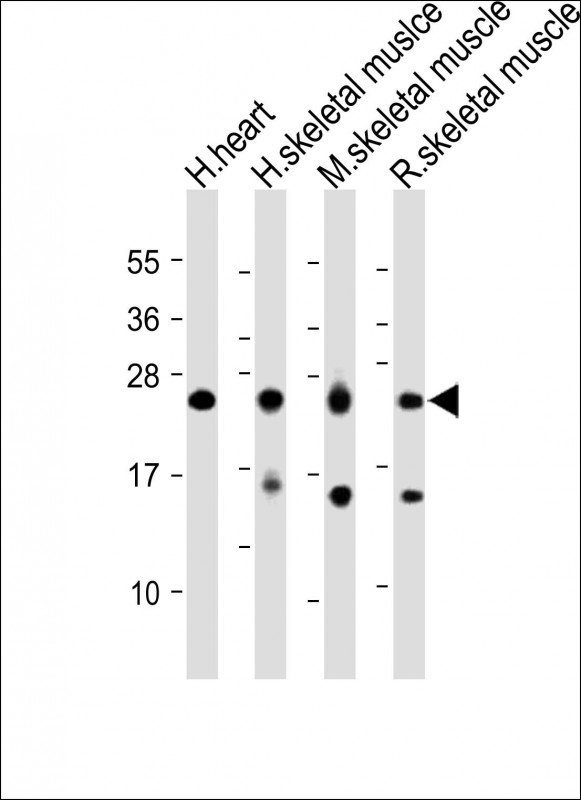
All lanes : Anti-MYL1 Antibody (Center) at 1:2000 dilution
Lane 1: Human heart lysate
Lane 2: Human skeletal muslce lysate
Lane 3: Mouse skeletal muscle lysate
Lane 4: Rat skeletal muscle lysate
Lysates/proteins at 20 µg per lane.
Secondary
Goat Anti-Rabbit IgG, (H+L), Peroxidase conjugated at 1/10000 dilution.
Predicted band size : 21 kDa
Blocking/Dilution buffer: 5% NFDM/TBST.
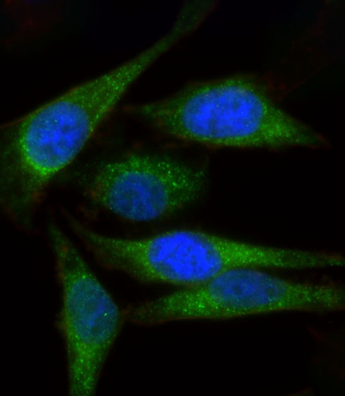
Immunofluorescent analysis of 4% paraformaldehyde-fixed, 0. 1% Triton X-100 permeabilized Hela cells labeling MYL1 with P34512 at 1/25 dilution, followed by Dylight® 488-conjugated goat anti-Rabbit IgG secondary antibody at 1/200 dilution (green). Immunofluorescence image showing Cytoplasm and Weak Nucleus staining on Hela cell line. The nuclear counter stain is DAPI (blue).
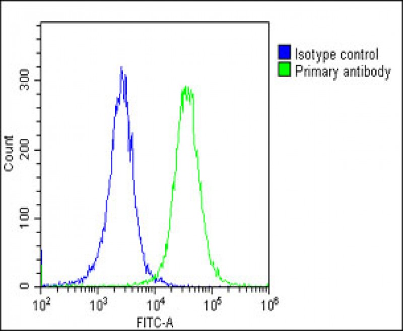
Overlay histogram showing Hela cells stained with P34512(green line). The cells were fixed with 2% paraformaldehyde (10 min) and then permeabilized with 90% methanol for 10 min. The cells were then icubated in 2% bovine serum albumin to block non-specific protein-protein interactions followed by the antibody (P34512, 1:25 dilution) for 60 min at 37ºC. The secondary antibody used was Goat-Anti-Rabbit IgG, DyLight® 488 Conjugated Highly Cross-Adsorbed(OE188374) at 1/200 dilution for 40 min at 37ºC. Isotype control antibody (blue line) was rabbit IgG1 (1μg/1x10^6 cells) used under the same conditions. Acquisition of >10, 000 events was performed.
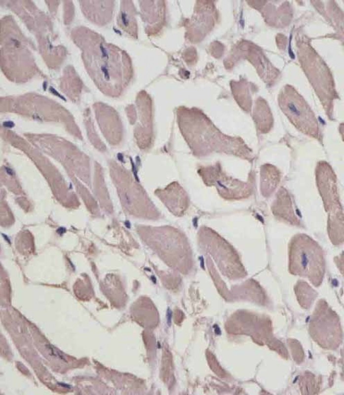
P34512 staining MYL1 in human heart tissue sections by Immunohistochemistry (IHC-P - paraformaldehyde-fixed, paraffin-embedded sections). Tissue was fixed with formaldehyde and blocked with 3% BSA for 0. 5 hour at room temperature; antigen retrieval was by heat mediation with a citrate buffer (pH6). Samples were incubated with primary antibody (1/25) for 1 hours at 37°C. A undiluted biotinylated goat polyvalent antibody was used as the secondary antibody.
Rabbit Polyclonal Antibody to MYL1
-
货号:
P34512 -
别名:
Myosin light chain 1/3, skeletal muscle isoform, MLC1/MLC3, MLC1F/MLC3F, Myosin light chain alkali 1/2, Myosin light chain A1/A2, MYL1 -
应用:
WB,IHC-P,IF,FCM -
反应种属:
Human, Mouse, Rat -
抗体类型:
Primary antibody -
Swissprot:
P05976 -
规格:
-
数量:
-+ -
说明书:

目录价¥1980


Rabbit Polyclonal Antibody to MYL1
Description |
|---|
Regulatory light chain of myosin. Does not bind calcium. |
Specification |
|
|---|---|
| Aliases | Myosin light chain 1/3, skeletal muscle isoform, MLC1/MLC3, MLC1F/MLC3F, Myosin light chain alkali 1/2, Myosin light chain A1/A2, MYL1 |
| Entrez GeneID | 4632 |
| Swissprot | P05976 |
| WB Predicted band size | 21.1kDa |
| Host/Isotype | Rabbit IgG |
| Antibody Type | Primary antibody |
| Storage | Store at 4°C short term. Aliquot and store at -20°C long term. Avoid freeze/thaw cycles. |
| Species Reactivity | Human, Mouse, Rat |
| Immunogen | This MYL1 antibody is generated from a rabbit immunized with a KLH conjugated synthetic peptide between 101-135 amino acids from the Central region of human MYL1. |
Application |
|
|---|---|
| WB | 1/2000 |
| IHC | 1/100-1/500 |
| IF/ICC | 1/25 |
| FCM | 1/25 |
Product Image
- All lanes : Anti-MYL1 Antibody (Center) at 1:2000 dilution Lane 1: Human heart lysate Lane 2: Human skeletal muslce lysate Lane 3: Mouse skeletal muscle lysate Lane 4: Rat skeletal muscle lysate Lysates/proteins at 20 µg per lane. Secondary Goat Anti-Rabbit IgG, (H+L), Peroxidase conjugated at 1/10000 dilution. Predicted band size : 21 kDa Blocking/Dilution buffer: 5% NFDM/TBST.
- Immunofluorescent analysis of 4% paraformaldehyde-fixed, 0. 1% Triton X-100 permeabilized Hela cells labeling MYL1 with P34512 at 1/25 dilution, followed by Dylight® 488-conjugated goat anti-Rabbit IgG secondary antibody at 1/200 dilution (green). Immunofluorescence image showing Cytoplasm and Weak Nucleus staining on Hela cell line. The nuclear counter stain is DAPI (blue).
- Overlay histogram showing Hela cells stained with P34512(green line). The cells were fixed with 2% paraformaldehyde (10 min) and then permeabilized with 90% methanol for 10 min. The cells were then icubated in 2% bovine serum albumin to block non-specific protein-protein interactions followed by the antibody (P34512, 1:25 dilution) for 60 min at 37ºC. The secondary antibody used was Goat-Anti-Rabbit IgG, DyLight® 488 Conjugated Highly Cross-Adsorbed(OE188374) at 1/200 dilution for 40 min at 37ºC. Isotype control antibody (blue line) was rabbit IgG1 (1μg/1x10^6 cells) used under the same conditions. Acquisition of >10, 000 events was performed.
- P34512 staining MYL1 in human heart tissue sections by Immunohistochemistry (IHC-P - paraformaldehyde-fixed, paraffin-embedded sections). Tissue was fixed with formaldehyde and blocked with 3% BSA for 0. 5 hour at room temperature; antigen retrieval was by heat mediation with a citrate buffer (pH6). Samples were incubated with primary antibody (1/25) for 1 hours at 37°C. A undiluted biotinylated goat polyvalent antibody was used as the secondary antibody.
For Reseach Only
Application Key:WB - Western Blot | IHC - Immunohistochemistry | ICC - Immunocytochemistry | FCM - Flow Cytometry | ELISA - Enzyme-linked Immunosorbent Assay | IP - Immunoprecipitation
#P34512

相关产品


联系方式
CONTACT-
联系电话:
0731-88388785 -
公司邮箱:
sales@promab.cn -
公司地址:
湖南省长沙市高新开发区林语路239号顺畅产业园5楼
 官方微信
官方微信
产品中心
Product技术服务
service客户留言
message