


提醒成功


 微信/QQ登录
搜索


 首页
首页
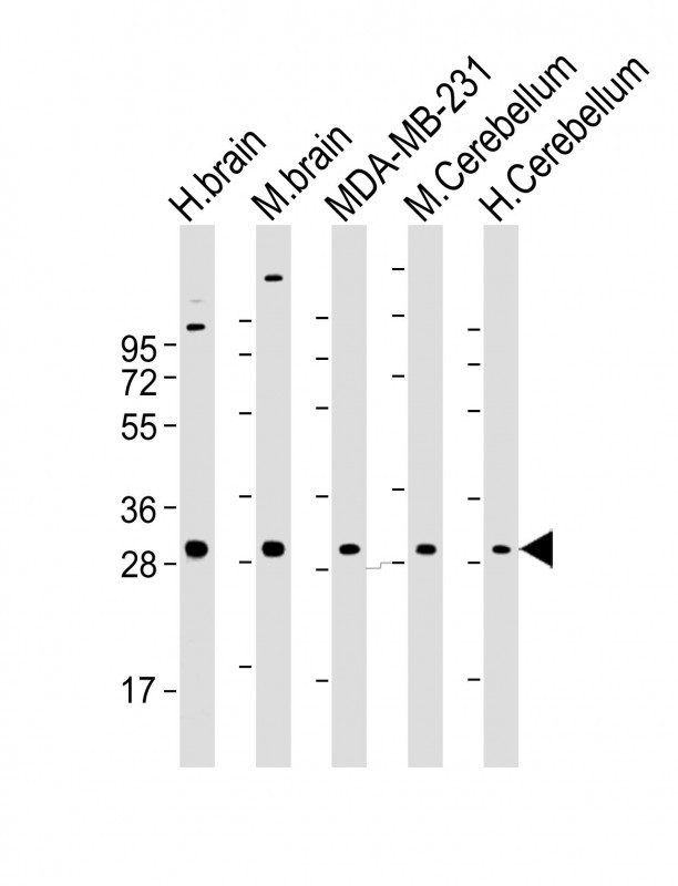
All lanes : Anti-RAB23 Antibody at 1:2000 dilution
Lane 1: human brain lysate
Lane 2: mouse brain lysate
Lane 3: MDA-MB-231 whole cell lysate
Lane 4: mouse Cerebellum lysate
Lane 5: human Cerebellum lysate
Lysates/proteins at 20 µg per lane.
Secondary
Goat Anti-mouse IgG, (H+L), Peroxidase conjugated at 1/10000 dilution.
Predicted band size : 27 kDa
Blocking/Dilution buffer: 5% NFDM/TBST.
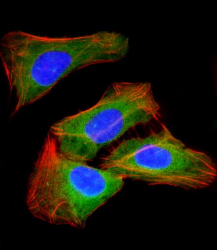
Immunofluorescent analysis of 4% paraformaldehyde-fixed, 0.1% Triton X-100 permeabilized U-2 OS (human osteosarcoma cell line) cells labeling RAB23 with AM2026a at 1/25 dilution, followed by Dylight® 488-conjugated goat anti-mouse IgG secondary antibody at 1/200 dilution (green). Immunofluorescence image showing cytoplasm staining on U-2 OS cell line. Cytoplasmic actin is detected with Dylight® 554 Phalloidin at 1/100 dilution (red).The nuclear counter stain is DAPI (blue).
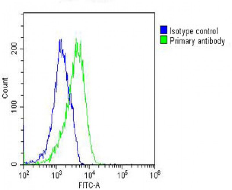
Overlay histogram showing U-2 OS cells stained with AM2026a(green line). The cells were fixed with 2% paraformaldehyde (10 min) and then permeabilized with 90% methanol for 10 min. The cells were then icubated in 2% bovine serum albumin to block non-specific protein-protein interactions followed by the antibody (AM2026a, 1:25 dilution) for 60 min at 37ºC. The secondary antibody used was Goat-Anti-Mouse IgG, DyLight® 488 Conjugated Highly Cross-Adsorbed(NH174309) at 1/200 dilution for 40 min at 37ºC. Isotype control antibody (blue line) was mouse IgG1(1μg/1x10^6 cells) used under the same conditions. Acquisition of >10, 000 events was performed.
Mouse Monoclonal Antibody to RAB23
-
货号:
P34372 -
别名:
Ras-related protein Rab-23, RAB23 -
应用:
WB,IF,FCM -
反应种属:
Human, Mouse -
抗体类型:
Primary antibody -
Swissprot:
Q9ULC3 -
规格:
-
数量:
-+ -
说明书:

目录价¥2180


Mouse Monoclonal Antibody to RAB23
Description |
|---|
The protein encoded by this gene belongs to the small GTPase superfamily, Rab family. It may be involved in small GTPase mediated signal transduction and intracellular protein transportation. Alternative splicing occurs at this locus and two transcript variants encoding the same protein have been identified. |
Specification |
|
|---|---|
| Aliases | Ras-related protein Rab-23, RAB23 |
| Entrez GeneID | 51715 |
| Swissprot | Q9ULC3 |
| WB Predicted band size | 26.7kDa |
| Host/Isotype | Mouse IgG1 |
| Antibody Type | Primary antibody |
| Storage | Store at 4°C short term. Aliquot and store at -20°C long term. Avoid freeze/thaw cycles. |
| Species Reactivity | Human, Mouse |
| Immunogen | Purified His-tagged RAB23 protein(Fragment) was used to produced this monoclonal antibody. |
| Formulation | Purified antibody in TBS with 0.05% sodium azide. |
Application |
|
|---|---|
| WB | 1/2000 |
| IF/ICC | 1/25 |
| FCM | 1/25 |
Product Image
- All lanes : Anti-RAB23 Antibody at 1:2000 dilution Lane 1: human brain lysate Lane 2: mouse brain lysate Lane 3: MDA-MB-231 whole cell lysate Lane 4: mouse Cerebellum lysate Lane 5: human Cerebellum lysate Lysates/proteins at 20 µg per lane. Secondary Goat Anti-mouse IgG, (H+L), Peroxidase conjugated at 1/10000 dilution. Predicted band size : 27 kDa Blocking/Dilution buffer: 5% NFDM/TBST.
- Immunofluorescent analysis of 4% paraformaldehyde-fixed, 0.1% Triton X-100 permeabilized U-2 OS (human osteosarcoma cell line) cells labeling RAB23 with AM2026a at 1/25 dilution, followed by Dylight® 488-conjugated goat anti-mouse IgG secondary antibody at 1/200 dilution (green). Immunofluorescence image showing cytoplasm staining on U-2 OS cell line. Cytoplasmic actin is detected with Dylight® 554 Phalloidin at 1/100 dilution (red).The nuclear counter stain is DAPI (blue).
- Overlay histogram showing U-2 OS cells stained with AM2026a(green line). The cells were fixed with 2% paraformaldehyde (10 min) and then permeabilized with 90% methanol for 10 min. The cells were then icubated in 2% bovine serum albumin to block non-specific protein-protein interactions followed by the antibody (AM2026a, 1:25 dilution) for 60 min at 37ºC. The secondary antibody used was Goat-Anti-Mouse IgG, DyLight® 488 Conjugated Highly Cross-Adsorbed(NH174309) at 1/200 dilution for 40 min at 37ºC. Isotype control antibody (blue line) was mouse IgG1(1μg/1x10^6 cells) used under the same conditions. Acquisition of >10, 000 events was performed.
For Reseach Only
Application Key:WB - Western Blot | IHC - Immunohistochemistry | ICC - Immunocytochemistry | FCM - Flow Cytometry | ELISA - Enzyme-linked Immunosorbent Assay | IP - Immunoprecipitation
#P34372

相关产品


联系方式
CONTACT-
联系电话:
0731-88388785 -
公司邮箱:
sales@promab.cn -
公司地址:
湖南省长沙市高新开发区林语路239号顺畅产业园5楼
 官方微信
官方微信
产品中心
Product技术服务
service客户留言
message