


提醒成功


 微信/QQ登录
搜索


 首页
首页
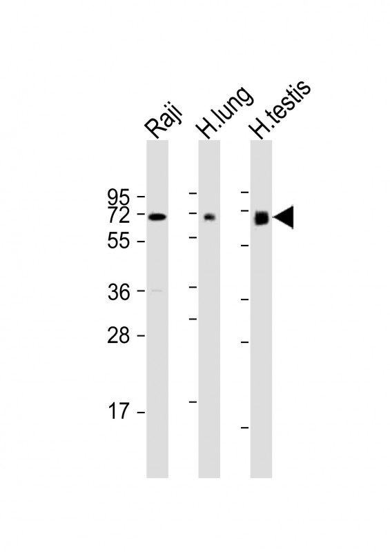
All lanes : Anti-ERVK-7 Antibody (N-Term) at 1:1000-1:2000 dilution
Lane 1: Raji whole cell lysate
Lane 2: human lung lysate
Lane 3: human testis lysate
Lysates/proteins at 20 µg per lane.
Secondary
Goat Anti-Rabbit IgG, (H+L), Peroxidase conjugated at 1/10000 dilution.
Predicted band size : 67 kDa
Blocking/Dilution buffer: 5% NFDM/TBST.
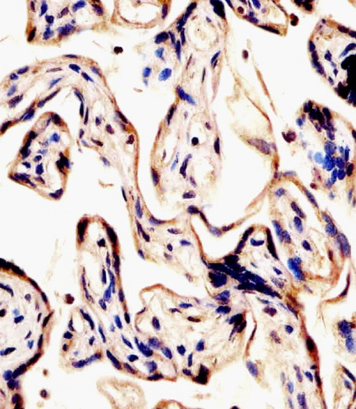
P33477 staining ERVK-7 in human placenta tissue sections by Immunohistochemistry (IHC-P - paraformaldehyde-fixed, paraffin-embedded sections). Tissue was fixed with formaldehyde and blocked with 3% BSA for 0. 5 hour at room temperature; antigen retrieval was by heat mediation with a citrate buffer (pH6). Samples were incubated with primary antibody (1/25) for 1 hours at 37°C. A undiluted biotinylated goat polyvalent antibody was used as the secondary antibody.
Rabbit Polyclonal Antibody to ERVK-7 (N-Term)
-
货号:
P33477 -
别名:
Endogenous retrovirus group K member 7 Env polyprotein, Envelope polyprotein, HERV-K(III) envelope protein, HERV-K102 envelope protein, HERV-K_1q22 provirus ancestral Env polyprotein, Surface protein, SU, Transmembrane protein, TM, ERVK-7 -
应用:
WB,IHC-P -
反应种属:
Human -
抗体类型:
Primary antibody -
Swissprot:
P61567 -
规格:
-
数量:
-+ -
说明书:

目录价¥1980


Rabbit Polyclonal Antibody to ERVK-7 (N-Term)
Description |
|---|
Retroviral envelope proteins mediate receptor recognition and membrane fusion during early infection. Endogenous envelope proteins may have kept, lost or modified their original function during evolution. TM anchors the envelope heterodimer to the viral membrane through one transmembrane domain. The other hydrophobic domain, called fusion peptide, mediates fusion of the viral membrane with the target cell membrane (By similarity). |
Specification |
|
|---|---|
| Aliases | Endogenous retrovirus group K member 7 Env polyprotein, Envelope polyprotein, HERV-K(III) envelope protein, HERV-K102 envelope protein, HERV-K_1q22 provirus ancestral Env polyprotein, Surface protein, SU, Transmembrane protein, TM, ERVK-7 |
| Swissprot | P61567 |
| WB Predicted band size | 66.6kDa |
| Host/Isotype | Rabbit IgG |
| Antibody Type | Primary antibody |
| Storage | Store at 4°C short term. Aliquot and store at -20°C long term. Avoid freeze/thaw cycles. |
| Species Reactivity | Human |
| Immunogen | This ERVK-7 antibody is generated from a rabbit immunized with a KLH conjugated synthetic peptide between 96-128 amino acids from human ERVK-7. |
Application |
|
|---|---|
| WB | 1/1000-1/2000 |
| IHC | 1/100-1/500 |
Product Image
- All lanes : Anti-ERVK-7 Antibody (N-Term) at 1:1000-1:2000 dilution Lane 1: Raji whole cell lysate Lane 2: human lung lysate Lane 3: human testis lysate Lysates/proteins at 20 µg per lane. Secondary Goat Anti-Rabbit IgG, (H+L), Peroxidase conjugated at 1/10000 dilution. Predicted band size : 67 kDa Blocking/Dilution buffer: 5% NFDM/TBST.
- P33477 staining ERVK-7 in human placenta tissue sections by Immunohistochemistry (IHC-P - paraformaldehyde-fixed, paraffin-embedded sections). Tissue was fixed with formaldehyde and blocked with 3% BSA for 0. 5 hour at room temperature; antigen retrieval was by heat mediation with a citrate buffer (pH6). Samples were incubated with primary antibody (1/25) for 1 hours at 37°C. A undiluted biotinylated goat polyvalent antibody was used as the secondary antibody.
For Reseach Only
Application Key:WB - Western Blot | IHC - Immunohistochemistry | ICC - Immunocytochemistry | FCM - Flow Cytometry | ELISA - Enzyme-linked Immunosorbent Assay | IP - Immunoprecipitation
#P33477

相关产品


联系方式
CONTACT-
联系电话:
0731-88388785 -
公司邮箱:
sales@promab.cn -
公司地址:
湖南省长沙市高新开发区林语路239号顺畅产业园5楼
 官方微信
官方微信
产品中心
Product技术服务
service客户留言
message