


提醒成功


 微信/QQ登录
搜索


 首页
首页
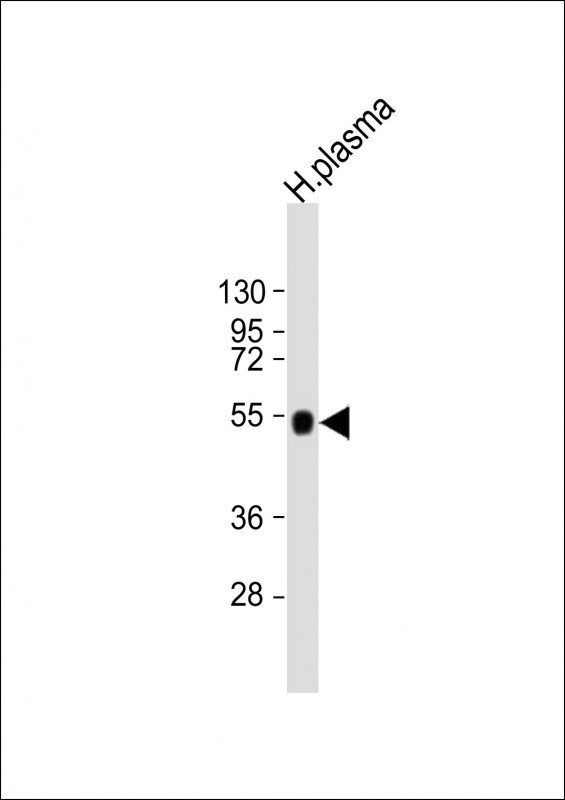
Anti-AHSG Antibody (C-term) at 1:2000 dilution + human plasma lysate
Lysates/proteins at 20 µg per lane.
Secondary
Goat Anti-Rabbit IgG, (H+L), Peroxidase conjugated at 1/10000 dilution.
Predicted band size : 39 kDa
Blocking/Dilution buffer: 5% NFDM/TBST.
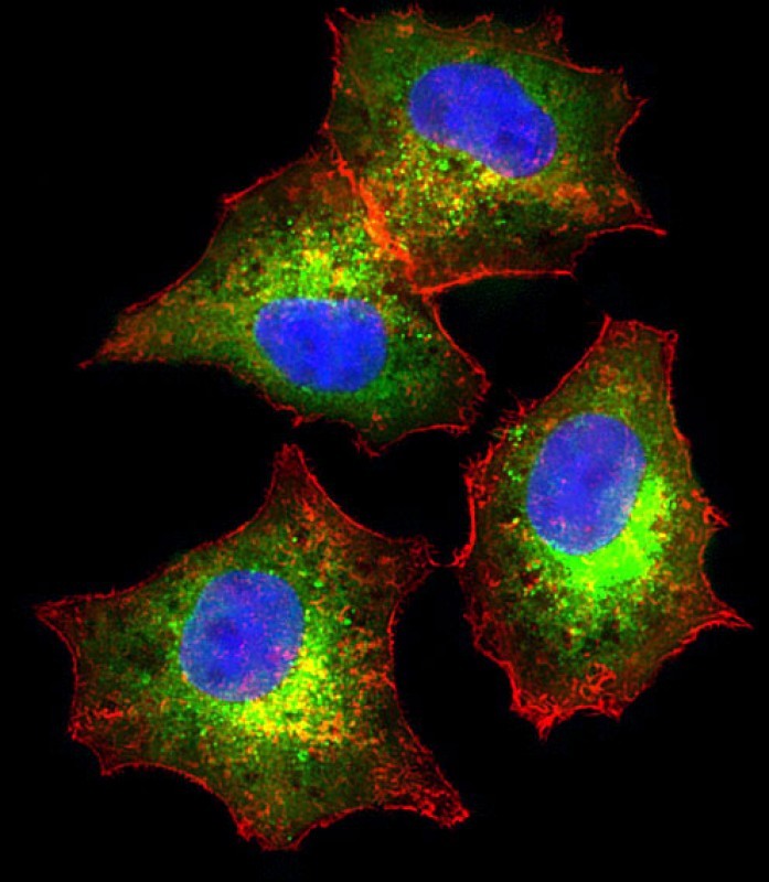
Immunofluorescent analysis of 4% paraformaldehyde-fixed, 0.1% Triton X-100 permeabilized HepG2 (human liver hepatocellular carcinoma cell line) cells labeling AHSG with P33383 at 1/25 dilution, followed by Dylight® 488-conjugated goat anti-rabbit IgG secondary antibody at 1/200 dilution (green). Immunofluorescence image showing cytoplasm staining on HepG2 cell line. Cytoplasmic actin is detected with Dylight® 554 Phalloidin at 1/100 dilution (red).The nuclear counter stain is DAPI (blue).
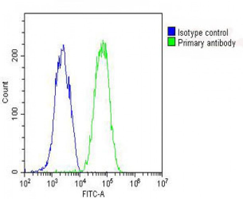
Overlay histogram showing HepG2 cells stained with P33383 (green line). The cells were fixed with 2% paraformaldehyde (10 min) and then permeabilized with 90% methanol for 10 min. The cells were then icubated in 2% bovine serum albumin to block non-specific protein-protein interactions followed by the antibody (P33383, 1:25 dilution) for 60 min at 37ºC. The secondary antibody used was Goat-Anti-Rabbit IgG, DyLight® 488 Conjugated Highly Cross-Adsorbed(OH191631) at 1/200 dilution for 40 min at 37ºC. Isotype control antibody (blue line) was rabbit IgG (1μg/1x10^6 cells) used under the same conditions. Acquisition of >10, 000 events was performed.
Rabbit Polyclonal Antibody to AHSG
-
货号:
P33383 -
别名:
Alpha-2-HS-glycoprotein, Alpha-2-Z-globulin, Ba-alpha-2-glycoprotein, Fetuin-A, Alpha-2-HS-glycoprotein chain A, Alpha-2-HS-glycoprotein chain B, AHSG, FETUA -
应用:
WB,IF,FCM -
反应种属:
Human -
抗体类型:
Primary antibody -
Swissprot:
P02765 -
规格:
-
数量:
-+ -
说明书:

目录价¥1980


Rabbit Polyclonal Antibody to AHSG
Description |
|---|
Alpha2-HS glycoprotein (AHSG), a glycoprotein present in the serum, is synthesized by hepatocytes. The AHSG molecule consists of two polypeptide chains, which are both cleaved from a proprotein encoded from a single mRNA. It is involved in several functions, such as endocytosis, brain development and the formation of bone tissue. The protein is commonly present in the cortical plate of the immature cerebral cortex and bone marrow hemopoietic matrix, and it has therefore been postulated that it participates in the development of the tissues. However, its exact significance is still obscure. |
Specification |
|
|---|---|
| Aliases | Alpha-2-HS-glycoprotein, Alpha-2-Z-globulin, Ba-alpha-2-glycoprotein, Fetuin-A, Alpha-2-HS-glycoprotein chain A, Alpha-2-HS-glycoprotein chain B, AHSG, FETUA |
| Entrez GeneID | 197 |
| Swissprot | P02765 |
| WB Predicted band size | 39.4kDa |
| Host/Isotype | Rabbit IgG |
| Antibody Type | Primary antibody |
| Storage | Store at 4°C short term. Aliquot and store at -20°C long term. Avoid freeze/thaw cycles. |
| Species Reactivity | Human |
| Immunogen | This AHSG antibody is generated from rabbits immunized with a KLH conjugated synthetic peptide between 247-276 amino acids from the C-terminal region of human AHSG. |
| Formulation | Purified antibody in PBS with 0.05% sodium azide. |
Application |
|
|---|---|
| WB | 1/2000 |
| IF/ICC | 1/25 |
| FCM | 1/25 |
Product Image
- Anti-AHSG Antibody (C-term) at 1:2000 dilution + human plasma lysate Lysates/proteins at 20 µg per lane. Secondary Goat Anti-Rabbit IgG, (H+L), Peroxidase conjugated at 1/10000 dilution. Predicted band size : 39 kDa Blocking/Dilution buffer: 5% NFDM/TBST.
- Immunofluorescent analysis of 4% paraformaldehyde-fixed, 0.1% Triton X-100 permeabilized HepG2 (human liver hepatocellular carcinoma cell line) cells labeling AHSG with P33383 at 1/25 dilution, followed by Dylight® 488-conjugated goat anti-rabbit IgG secondary antibody at 1/200 dilution (green). Immunofluorescence image showing cytoplasm staining on HepG2 cell line. Cytoplasmic actin is detected with Dylight® 554 Phalloidin at 1/100 dilution (red).The nuclear counter stain is DAPI (blue).
- Overlay histogram showing HepG2 cells stained with P33383 (green line). The cells were fixed with 2% paraformaldehyde (10 min) and then permeabilized with 90% methanol for 10 min. The cells were then icubated in 2% bovine serum albumin to block non-specific protein-protein interactions followed by the antibody (P33383, 1:25 dilution) for 60 min at 37ºC. The secondary antibody used was Goat-Anti-Rabbit IgG, DyLight® 488 Conjugated Highly Cross-Adsorbed(OH191631) at 1/200 dilution for 40 min at 37ºC. Isotype control antibody (blue line) was rabbit IgG (1μg/1x10^6 cells) used under the same conditions. Acquisition of >10, 000 events was performed.
For Reseach Only
Application Key:WB - Western Blot | IHC - Immunohistochemistry | ICC - Immunocytochemistry | FCM - Flow Cytometry | ELISA - Enzyme-linked Immunosorbent Assay | IP - Immunoprecipitation
#P33383

相关产品


联系方式
CONTACT-
联系电话:
0731-88388785 -
公司邮箱:
sales@promab.cn -
公司地址:
湖南省长沙市高新开发区林语路239号顺畅产业园5楼
 官方微信
官方微信
产品中心
Product技术服务
service客户留言
message