


提醒成功


 微信/QQ登录
搜索


 首页
首页
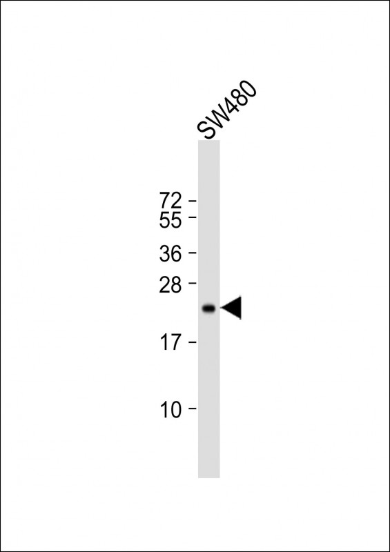
Anti-TIMP2 Antibody at 1:500 dilution + SW480 whole cell lysate
Lysates/proteins at 20 µg per lane.
Secondary
Goat Anti-mouse IgG, (H+L), Peroxidase conjugated at 1/10000 dilution.
Predicted band size : 24 kDa
Blocking/Dilution buffer: 5% NFDM/TBST.
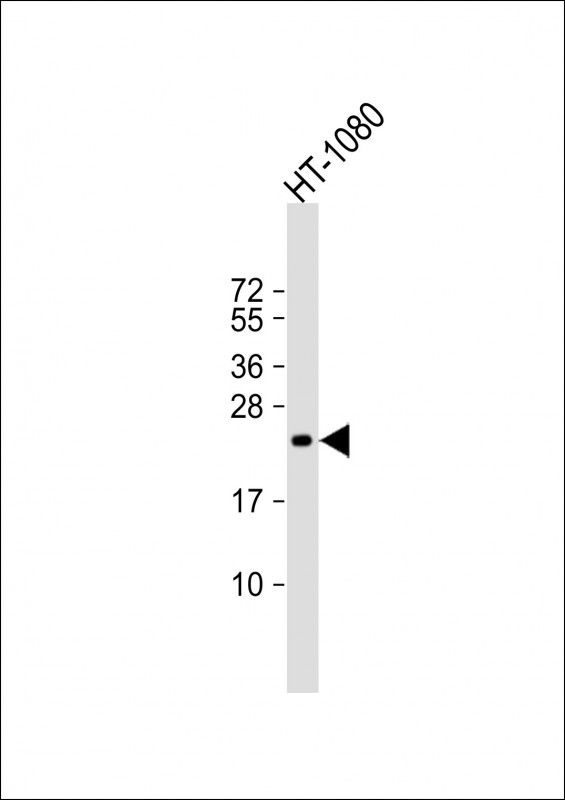
Anti-TIMP2 Antibody at 1:2000 dilution + HT-1080 whole cell lysate
Lysates/proteins at 20 µg per lane.
Secondary
Goat Anti-mouse IgG, (H+L), Peroxidase conjugated at 1/10000 dilution.
Predicted band size : 24 kDa
Blocking/Dilution buffer: 5% NFDM/TBST.
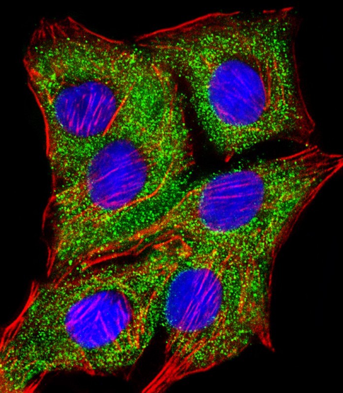
Immunofluorescent analysis of 4% paraformaldehyde-fixed, 0.1% Triton X-100 permeabilized A549 (human lung adenocarcinoma epithelial cell line) cells labeling TIMP2 with P33380 at 1/25 dilution, followed by Dylight® 488-conjugated goat anti-mouse IgG secondary antibody at 1/200 dilution (green). Immunofluorescence image showing cytoplasm staining on A549 cell line. Cytoplasmic actin is detected with Dylight® 554 Phalloidin at 1/100 dilution (red).The nuclear counter stain is DAPI (blue).
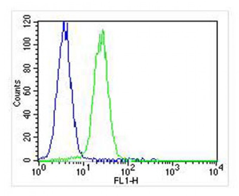
Overlay histogram showing K562 cells stained with P33380 (green line). The cells were fixed with 2% paraformaldehyde (10 min) and then permeabilized with 90% methanol for 10 min. The cells were then icubated in 2% bovine serum albumin to block non-specific protein-protein interactions followed by the antibody (P33380, 1:25 dilution) for 60 min at 37ºC. The secondary antibody used was Goat-Anti-Mouse IgG, DyLight® 488 Conjugated Highly Cross-Adsorbed(NA168821) at 1/400 dilution for 40 min at 37ºC. Isotype control antibody (blue line) was mouse IgG1 (1μg/1x10^6 cells) used under the same conditions. Acquisition of >10, 000 events was performed.
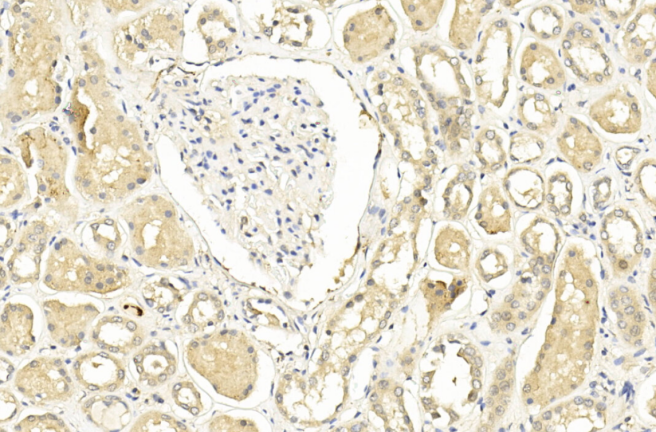
Immunohistochemical analysis of paraffin-embedded Human kidney section using Pink1(Cat#P33380). P33380 was diluted at 1:200 dilution. A undiluted biotinylated goat polyvalent antibody was used as the secondary, followed by DAB staining.
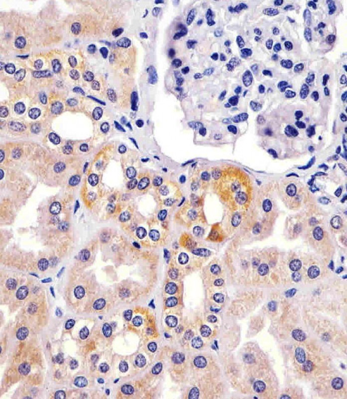
P33380 staining TIMP2 in human kidney sections by Immunohistochemistry (IHC-P - paraformaldehyde-fixed, paraffin-embedded sections). Tissue was fixed with formaldehyde and blocked with 3% BSA for 0. 5 hour at room temperature; antigen retrieval was by heat mediation with a citrate buffer (pH6). Samples were incubated with primary antibody (1/25) for 1 hours at 37°C. A undiluted biotinylated goat polyvalent antibody was used as the secondary antibody.
Mouse Monoclonal Antibody to TIMP2
-
货号:
P33380 -
别名:
Metalloproteinase inhibitor 2, CSC-21K, Tissue inhibitor of metalloproteinases 2, TIMP-2, TIMP2 -
应用:
WB,IHC-P,IF,FCM -
反应种属:
Human, Mouse, Rat -
抗体类型:
Primary antibody -
Swissprot:
P16035 -
规格:
-
数量:
-+ -
说明书:

目录价¥2180


Mouse Monoclonal Antibody to TIMP2
Description |
|---|
Complexes with metalloproteinases (such as collagenases) and irreversibly inactivates them by binding to their catalytic zinc cofactor. Known to act on MMP-1, MMP-2, MMP-3, MMP-7, MMP-8, MMP-9, MMP-10, MMP-13, MMP-14, MMP-15, MMP-16 and MMP-19. |
Specification |
|
|---|---|
| Aliases | Metalloproteinase inhibitor 2, CSC-21K, Tissue inhibitor of metalloproteinases 2, TIMP-2, TIMP2 |
| Entrez GeneID | 7077 |
| Swissprot | P16035 |
| WB Predicted band size | 24.4kDa |
| Host/Isotype | Mouse IgG1 |
| Antibody Type | Primary antibody |
| Storage | Store at 4°C short term. Aliquot and store at -20°C long term. Avoid freeze/thaw cycles. |
| Species Reactivity | Human, Mouse, Rat |
| Immunogen | This TIMP2 antibody is generated from a mouse immunized with arecombinant protein of human TIMP2. |
Application |
|
|---|---|
| WB | 1/500-1/2000 |
| IHC | 1/100-1/500 |
| IF/ICC | 1/25 |
| FCM | 1/25 |
Product Image
- Anti-TIMP2 Antibody at 1:500 dilution + SW480 whole cell lysate Lysates/proteins at 20 µg per lane. Secondary Goat Anti-mouse IgG, (H+L), Peroxidase conjugated at 1/10000 dilution. Predicted band size : 24 kDa Blocking/Dilution buffer: 5% NFDM/TBST.
- Anti-TIMP2 Antibody at 1:2000 dilution + HT-1080 whole cell lysate Lysates/proteins at 20 µg per lane. Secondary Goat Anti-mouse IgG, (H+L), Peroxidase conjugated at 1/10000 dilution. Predicted band size : 24 kDa Blocking/Dilution buffer: 5% NFDM/TBST.
- Immunofluorescent analysis of 4% paraformaldehyde-fixed, 0.1% Triton X-100 permeabilized A549 (human lung adenocarcinoma epithelial cell line) cells labeling TIMP2 with P33380 at 1/25 dilution, followed by Dylight® 488-conjugated goat anti-mouse IgG secondary antibody at 1/200 dilution (green). Immunofluorescence image showing cytoplasm staining on A549 cell line. Cytoplasmic actin is detected with Dylight® 554 Phalloidin at 1/100 dilution (red).The nuclear counter stain is DAPI (blue).
- Overlay histogram showing K562 cells stained with P33380 (green line). The cells were fixed with 2% paraformaldehyde (10 min) and then permeabilized with 90% methanol for 10 min. The cells were then icubated in 2% bovine serum albumin to block non-specific protein-protein interactions followed by the antibody (P33380, 1:25 dilution) for 60 min at 37ºC. The secondary antibody used was Goat-Anti-Mouse IgG, DyLight® 488 Conjugated Highly Cross-Adsorbed(NA168821) at 1/400 dilution for 40 min at 37ºC. Isotype control antibody (blue line) was mouse IgG1 (1μg/1x10^6 cells) used under the same conditions. Acquisition of >10, 000 events was performed.
- Immunohistochemical analysis of paraffin-embedded Human kidney section using Pink1(Cat#P33380). P33380 was diluted at 1:200 dilution. A undiluted biotinylated goat polyvalent antibody was used as the secondary, followed by DAB staining.
- P33380 staining TIMP2 in human kidney sections by Immunohistochemistry (IHC-P - paraformaldehyde-fixed, paraffin-embedded sections). Tissue was fixed with formaldehyde and blocked with 3% BSA for 0. 5 hour at room temperature; antigen retrieval was by heat mediation with a citrate buffer (pH6). Samples were incubated with primary antibody (1/25) for 1 hours at 37°C. A undiluted biotinylated goat polyvalent antibody was used as the secondary antibody.
For Reseach Only
Application Key:WB - Western Blot | IHC - Immunohistochemistry | ICC - Immunocytochemistry | FCM - Flow Cytometry | ELISA - Enzyme-linked Immunosorbent Assay | IP - Immunoprecipitation
#P33380

相关产品


联系方式
CONTACT-
联系电话:
0731-88388785 -
公司邮箱:
sales@promab.cn -
公司地址:
湖南省长沙市高新开发区林语路239号顺畅产业园5楼
 官方微信
官方微信
产品中心
Product技术服务
service客户留言
message