


提醒成功


 微信/QQ登录
搜索


 首页
首页
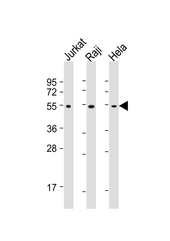
All lanes : Anti-XIAP Antibody at 1:1000-1:4000 dilution
Lane 1: Jurkat whole cell lysates
Lane 2: Raji whole cell lysates
Lane 3: Hela whole cell lysates
Lysates/proteins at 20 μg per lane.
Secondary
Goat Anti-mouse IgG, (H+L), Peroxidase conjugated at 1/10000 dilution.
Predicted band size : 57 kDa
Blocking/Dilution buffer: 5% NFDM/TBST.
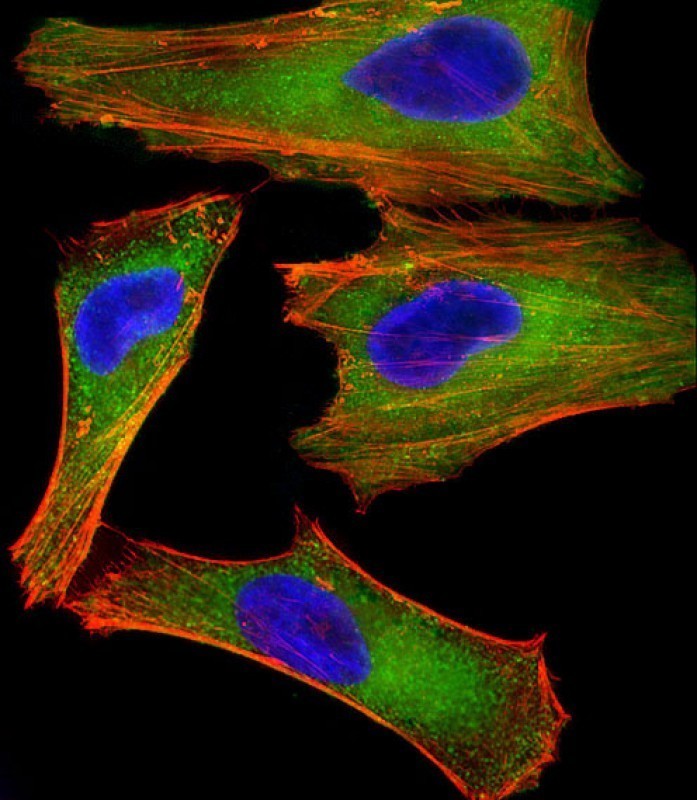
Immunofluorescent analysis of 4% paraformaldehyde-fixed, 0.1% Triton X-100 permeabilized HeLa (human cervical epithelial adenocarcinoma cell line) cells labeling XIAP with P33213 at 1/25 dilution, followed by Dylight® 488-conjugated goat anti-mouse IgG secondary antibody at 1/200 dilution (green). Immunofluorescence image showing cytoplasm staining on HeLa cell line. Cytoplasmic actin is detected with Dylight® 554 Phalloidin at 1/100 dilution (red).The nuclear counter stain is DAPI (blue).
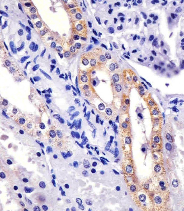
P33213 staining XIAP in human kidney sections by Immunohistochemistry (IHC-P - paraformaldehyde-fixed, paraffin-embedded sections). Tissue was fixed with formaldehyde and blocked with 3% BSA for 0. 5 hour at room temperature; antigen retrieval was by heat mediation with a citrate buffer (pH6). Samples were incubated with primary antibody (1/25) for 1 hours at 37°C. A undiluted biotinylated goat polyvalent antibody was used as the secondary antibody.
Mouse Monoclonal Antibody to XIAP
-
货号:
P33213 -
别名:
E3 ubiquitin-protein ligase XIAP, 632-, Baculoviral IAP repeat-containing protein 4, IAP-like protein, ILP, hILP, Inhibitor of apoptosis protein 3, IAP-3, hIAP-3, hIAP3, X-linked inhibitor of apoptosis protein, X-linked IAP, XIAP, API3, BIRC4, IAP3 -
应用:
WB,IHC-P,IF -
反应种属:
Human, Mouse, Rat -
抗体类型:
Primary antibody -
Swissprot:
P98170 -
规格:
-
数量:
-+ -
说明书:

目录价¥2180


Mouse Monoclonal Antibody to XIAP
Description |
|---|
Multi-functional protein which regulates not only caspases and apoptosis, but also modulates inflammatory signaling and immunity, copper homeostasis, mitogenic kinase signaling, cell proliferation, as well as cell invasion and metastasis. Acts as a direct caspase inhibitor. Directly bind to the active site pocket of CASP3 and CASP7 and obstructs substrate entry. Inactivates CASP9 by keeping it in a monomeric, inactive state. Acts as an E3 ubiquitin-protein ligase regulating NF-kappa-B signaling and the target proteins for its E3 ubiquitin-protein ligase activity include: RIPK1, CASP3, CASP7, CASP8, CASP9, MAP3K2/MEKK2, DIABLO/SMAC, AIFM1, CCS and BIRC5/survivin. Ubiquitinion of CCS leads to enhancement of its chaperone activity toward its physiologic target, SOD1, rather than proteasomal degradation. Ubiquitinion of MAP3K2/MEKK2 and AIFM1 does not lead to proteasomal degradation. Plays a role in copper homeostasis by ubiquitinationg COMMD1 and promoting its proteasomal degradation. Can also function as E3 ubiquitin-protein ligase of the NEDD8 conjugation pathway, targeting effector caspases for neddylation and inactivation. Regulates the BMP signaling pathway and the SMAD and MAP3K7/TAK1 dependent pathways leading to NF-kappa-B and JNK activation. Acts as an important regulator of innate immune signaling via regulation of Nodlike receptors (NLRs). Protects cells from spontaneous formation of the ripoptosome, a large multi-protein complex that has the capability to kill cancer cells in a caspase-dependent and caspase-independent manner. Suppresses ripoptosome formation by ubiquitinating RIPK1 and CASP8. Acts as a positive regulator of Wnt signaling and ubiquitinates TLE1, TLE2, TLE3, TLE4 and AES. Ubiquitination of TLE3 results in inhibition of its interaction with TCF7L2/TCF4 thereby allowing efficient recruitment and binding of the transcriptional coactivator beta- catenin to TCF7L2/TCF4 that is required to initiate a Wnt-specific transcriptional program. |
Specification |
|
|---|---|
| Aliases | E3 ubiquitin-protein ligase XIAP, 632-, Baculoviral IAP repeat-containing protein 4, IAP-like protein, ILP, hILP, Inhibitor of apoptosis protein 3, IAP-3, hIAP-3, hIAP3, X-linked inhibitor of apoptosis protein, X-linked IAP, XIAP, API3, BIRC4, IAP3 |
| Entrez GeneID | 331 |
| Swissprot | P98170 |
| WB Predicted band size | 56.7kDa |
| Host/Isotype | Mouse IgG1 |
| Antibody Type | Primary antibody |
| Storage | Store at 4°C short term. Aliquot and store at -20°C long term. Avoid freeze/thaw cycles. |
| Species Reactivity | Human, Mouse, Rat |
| Immunogen | This XIAP antibody is generated from a mouse immunized with a recombinant protein human XIAP . |
Application |
|
|---|---|
| WB | 1/1000-1/4000 |
| IHC | 1/100-1/500 |
| IF/ICC | 1/25 |
Product Image
- All lanes : Anti-XIAP Antibody at 1:1000-1:4000 dilution Lane 1: Jurkat whole cell lysates Lane 2: Raji whole cell lysates Lane 3: Hela whole cell lysates Lysates/proteins at 20 μg per lane. Secondary Goat Anti-mouse IgG, (H+L), Peroxidase conjugated at 1/10000 dilution. Predicted band size : 57 kDa Blocking/Dilution buffer: 5% NFDM/TBST.
- Immunofluorescent analysis of 4% paraformaldehyde-fixed, 0.1% Triton X-100 permeabilized HeLa (human cervical epithelial adenocarcinoma cell line) cells labeling XIAP with P33213 at 1/25 dilution, followed by Dylight® 488-conjugated goat anti-mouse IgG secondary antibody at 1/200 dilution (green). Immunofluorescence image showing cytoplasm staining on HeLa cell line. Cytoplasmic actin is detected with Dylight® 554 Phalloidin at 1/100 dilution (red).The nuclear counter stain is DAPI (blue).
- P33213 staining XIAP in human kidney sections by Immunohistochemistry (IHC-P - paraformaldehyde-fixed, paraffin-embedded sections). Tissue was fixed with formaldehyde and blocked with 3% BSA for 0. 5 hour at room temperature; antigen retrieval was by heat mediation with a citrate buffer (pH6). Samples were incubated with primary antibody (1/25) for 1 hours at 37°C. A undiluted biotinylated goat polyvalent antibody was used as the secondary antibody.
For Reseach Only
Application Key:WB - Western Blot | IHC - Immunohistochemistry | ICC - Immunocytochemistry | FCM - Flow Cytometry | ELISA - Enzyme-linked Immunosorbent Assay | IP - Immunoprecipitation
#P33213

相关产品


联系方式
CONTACT-
联系电话:
0731-88388785 -
公司邮箱:
sales@promab.cn -
公司地址:
湖南省长沙市高新开发区林语路239号顺畅产业园5楼
 官方微信
官方微信
产品中心
Product技术服务
service客户留言
message