


提醒成功


 微信/QQ登录
搜索


 首页
首页
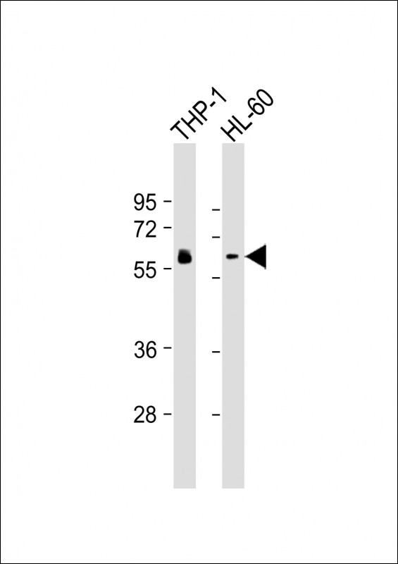
All lanes : Anti-HCK Antibody at 1:2000-1:4000 dilution
Lane 1: THP-1 whole cell lysates
Lane 2: HL-60 whole cell lysates
Lysates/proteins at 20 μg per lane.
Secondary
Goat Anti-mouse IgG, (H+L), Peroxidase conjugated at 1/10000 dilution.
Predicted band size : 60 kDa
Blocking/Dilution buffer: 5% NFDM/TBST.
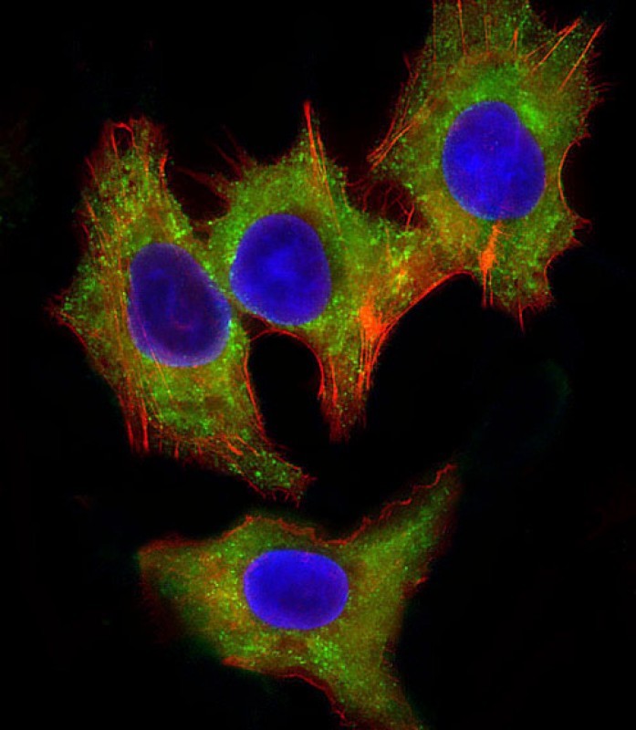
Immunofluorescent analysis of 4% paraformaldehyde-fixed, 0.1% Triton X-100 permeabilized HepG2 (human liver hepatocellular carcinoma cell line) cells labeling HCK with P33212 at 1/25 dilution, followed by Dylight® 488-conjugated goat anti-mouse IgG secondary antibody at 1/200 dilution (green). Immunofluorescence image showing cytoplasm staining on HepG2 cell line. Cytoplasmic actin is detected with Dylight® 554 Phalloidin at 1/100 dilution (red).The nuclear counter stain is DAPI (blue).
Mouse Monoclonal Antibody to HCK
-
货号:
P33212 -
别名:
Tyrosine-protein kinase HCK, Hematopoietic cell kinase, Hemopoietic cell kinase, p59-HCK/p60-HCK, p59Hck, p61Hck, HCK -
应用:
WB,IF -
反应种属:
Human, Mouse, Rat -
抗体类型:
Primary antibody -
Swissprot:
P08631 -
规格:
-
数量:
-+ -
说明书:

目录价¥2180


Mouse Monoclonal Antibody to HCK
Description |
|---|
Non-receptor tyrosine-protein kinase found in hematopoietic cells that transmits signals from cell surface receptors and plays an important role in the regulation of innate immune responses, including neutrophil, monocyte, macrophage and mast cell functions, phagocytosis, cell survival and proliferation, cell adhesion and migration. Acts downstream of receptors that bind the Fc region of immunoglobulins, such as FCGR1A and FCGR2A, but also CSF3R, PLAUR, the receptors for IFNG, IL2, IL6 and IL8, and integrins, such as ITGB1 and ITGB2. During the phagocytic process, mediates mobilization of secretory lysosomes, degranulation, and activation of NADPH oxidase to bring about the respiratory burst. Plays a role in the release of inflammatory molecules. Promotes reorganization of the actin cytoskeleton and actin polymerization, formation of podosomes and cell protrusions. Inhibits TP73-mediated transcription activation and TP73-mediated apoptosis. Phosphorylates CBL in response to activation of immunoglobulin gamma Fc region receptors. Phosphorylates ADAM15, BCR, ELMO1, FCGR2A, GAB1, GAB2, RAPGEF1, STAT5B, TP73, VAV1 and WAS. |
Specification |
|
|---|---|
| Aliases | Tyrosine-protein kinase HCK, Hematopoietic cell kinase, Hemopoietic cell kinase, p59-HCK/p60-HCK, p59Hck, p61Hck, HCK |
| Entrez GeneID | 3055 |
| Swissprot | P08631 |
| WB Predicted band size | 59.6kDa |
| Host/Isotype | Mouse IgG1 |
| Antibody Type | Primary antibody |
| Storage | Store at 4°C short term. Aliquot and store at -20°C long term. Avoid freeze/thaw cycles. |
| Species Reactivity | Human, Mouse, Rat |
| Immunogen | This HCK antibody is generated from a mouse immunized with a recombinant protein of human HCK. |
Application |
|
|---|---|
| WB | 1/2002-1/4000 |
| IF/ICC | 1/25 |
Product Image
- All lanes : Anti-HCK Antibody at 1:2000-1:4000 dilution Lane 1: THP-1 whole cell lysates Lane 2: HL-60 whole cell lysates Lysates/proteins at 20 μg per lane. Secondary Goat Anti-mouse IgG, (H+L), Peroxidase conjugated at 1/10000 dilution. Predicted band size : 60 kDa Blocking/Dilution buffer: 5% NFDM/TBST.
- Immunofluorescent analysis of 4% paraformaldehyde-fixed, 0.1% Triton X-100 permeabilized HepG2 (human liver hepatocellular carcinoma cell line) cells labeling HCK with P33212 at 1/25 dilution, followed by Dylight® 488-conjugated goat anti-mouse IgG secondary antibody at 1/200 dilution (green). Immunofluorescence image showing cytoplasm staining on HepG2 cell line. Cytoplasmic actin is detected with Dylight® 554 Phalloidin at 1/100 dilution (red).The nuclear counter stain is DAPI (blue).
For Reseach Only
Application Key:WB - Western Blot | IHC - Immunohistochemistry | ICC - Immunocytochemistry | FCM - Flow Cytometry | ELISA - Enzyme-linked Immunosorbent Assay | IP - Immunoprecipitation
#P33212

相关产品


联系方式
CONTACT-
联系电话:
0731-88388785 -
公司邮箱:
sales@promab.cn -
公司地址:
湖南省长沙市高新开发区林语路239号顺畅产业园5楼
 官方微信
官方微信
产品中心
Product技术服务
service客户留言
message