


提醒成功


 微信/QQ登录
搜索


 首页
首页
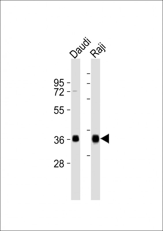
All lanes : Anti-HLA-DQA1 Antibody (C-term) at 1:8000 dilution
Lane 1: Daudi whole cell lysates
Lane 2: Raji whole cell lysates
Lysates/proteins at 20 µg per lane.
Secondary
Goat Anti-Rabbit IgG, (H+L), Peroxidase conjugated at 1/10000 dilution
Predicted band size : 28 kDa
Blocking/Dilution buffer: 5% NFDM/TBST.
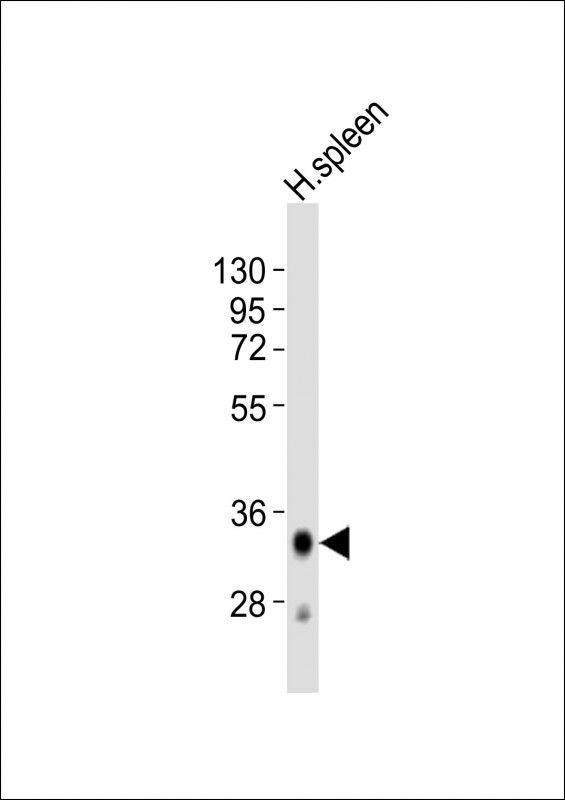
Anti-HLA-DQA1 Antibody (C-term)at 1:2000 dilution + human spleen lysates
Lysates/proteins at 20 µg per lane.
Secondary
Goat Anti-Rabbit IgG, (H+L), Peroxidase conjugated at 1/10000 dilution
Predicted band size : 28 kDa
Blocking/Dilution buffer: 5% NFDM/TBST.
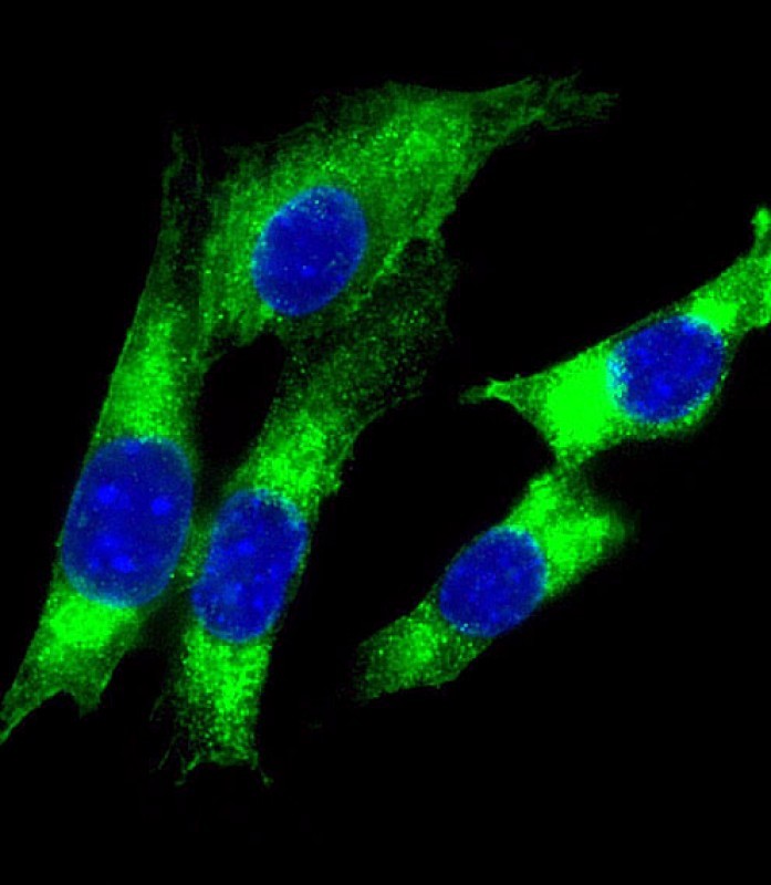
Immunofluorescent analysis of 4% paraformaldehyde-fixed, 0.1% Triton X-100 permeabilized NIH/3T3 (mouse embryonic fibroblast cell line) cells labeling HLA-DQA1 with P33168 at 1/25 dilution, followed by Dylight® 488-conjugated goat anti-rabbit IgG secondary antibody at 1/200 dilution (green). Immunofluorescence image showing cytoplasm staining on NIH/3T3 cell line. The nuclear counter stain is DAPI (blue).
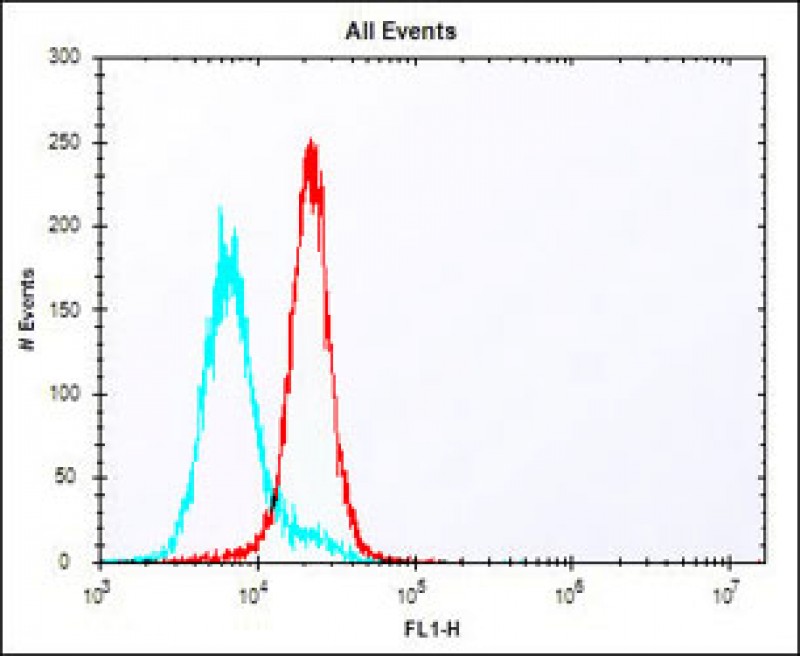
Overlay histogram showing K562 cells stained with P33168 (red line). The cells were fixed with 2% paraformaldehyde (10 min) and then permeabilized with 90% methanol for 10 min. The cells were then icubated in 2% bovine serum albumin to block non-specific protein-protein interactions followed by the antibody (P33168, 1:25 dilution) for 60 min at 37ºC. The secondary antibody used was Alexa Fluor® 488 goat anti-rabbit lgG (H+L) (1583138) at 1/400 dilution for 40 min at 37ºC. Isotype control antibody (blue line) was rabbit IgG1 (1μg/1x10^6 cells) used under the same conditions. Acquisition of >10, 000 events was performed.
Rabbit Polyclonal Antibody to HLA-DQA1
-
货号:
P33168 -
别名:
HLA class II histocompatibility antigen, DQ alpha 1 chain, DC-1 alpha chain, DC-alpha, HLA-DCA, MHC class II DQA1, HLA-DQA1 -
应用:
WB,IF,FCM -
反应种属:
Human, Mouse -
抗体类型:
Primary antibody -
Swissprot:
P01909 -
规格:
-
数量:
-+ -
说明书:

目录价¥1980


Rabbit Polyclonal Antibody to HLA-DQA1
Description |
|---|
Binds peptides derived from antigens that access the endocytic route of antigen presenting cells (APC) and presents them on the cell surface for recognition by the CD4 T-cells. The peptide binding cleft accommodates peptides of 10-30 residues. The peptides presented by MHC class II molecules are generated mostly by degradation of proteins that access the endocytic route, where they are processed by lysosomal proteases and other hydrolases. Exogenous antigens that have been endocytosed by the APC are thus readily available for presentation via MHC II molecules, and for this reason this antigen presentation pathway is usually referred to as exogenous. As membrane proteins on their way to degradation in lysosomes as part of their normal turn-over are also contained in the endosomal/lysosomal compartments, exogenous antigens must compete with those derived from endogenous components. Autophagy is also a source of endogenous peptides, autophagosomes constitutively fuse with MHC class II loading compartments. In addition to APCs, other cells of the gastrointestinal tract, such as epithelial cells, express MHC class II molecules and CD74 and act as APCs, which is an unusual trait of the GI tract. To produce a MHC class II molecule that presents an antigen, three MHC class II molecules (heterodimers of an alpha and a beta chain) associate with a CD74 trimer in the ER to form a heterononamer. Soon after the entry of this complex into the endosomal/lysosomal system where antigen processing occurs, CD74 undergoes a sequential degradation by various proteases, including CTSS and CTSL, leaving a small fragment termed CLIP (class-II-associated invariant chain peptide). The removal of CLIP is facilitated by HLA-DM via direct binding to the alpha-beta-CLIP complex so that CLIP is released. HLA-DM stabilizes MHC class II molecules until primary high affinity antigenic peptides are bound. The MHC II molecule bound to a peptide is then transported to the cell membrane surface. In B-cells, the interaction between HLA-DM and MHC class II molecules is regulated by HLA-DO. Primary dendritic cells (DCs) also to express HLA-DO. Lysosomal microenvironment has been implicated in the regulation of antigen loading into MHC II molecules, increased acidification produces increased proteolysis and efficient peptide loading. |
Specification |
|
|---|---|
| Aliases | HLA class II histocompatibility antigen, DQ alpha 1 chain, DC-1 alpha chain, DC-alpha, HLA-DCA, MHC class II DQA1, HLA-DQA1 |
| Entrez GeneID | 3117 |
| Swissprot | P01909 |
| WB Predicted band size | 27.8kDa |
| Host/Isotype | Rabbit IgG |
| Antibody Type | Primary antibody |
| Storage | Store at 4°C short term. Aliquot and store at -20°C long term. Avoid freeze/thaw cycles. |
| Species Reactivity | Human, Mouse |
| Immunogen | This HLA-DQA1 antibody is generated from a rabbit immunized with a KLH conjugated synthetic peptide between 188-222 amino acids from the C-terminal region of human HLA-DQA1. |
Application |
|
|---|---|
| WB | 1/2000-1/8000 |
| IF/ICC | 1/25 |
| FCM | 1/25 |
Product Image
- All lanes : Anti-HLA-DQA1 Antibody (C-term) at 1:8000 dilution Lane 1: Daudi whole cell lysates Lane 2: Raji whole cell lysates Lysates/proteins at 20 µg per lane. Secondary Goat Anti-Rabbit IgG, (H+L), Peroxidase conjugated at 1/10000 dilution Predicted band size : 28 kDa Blocking/Dilution buffer: 5% NFDM/TBST.
- Anti-HLA-DQA1 Antibody (C-term)at 1:2000 dilution + human spleen lysates Lysates/proteins at 20 µg per lane. Secondary Goat Anti-Rabbit IgG, (H+L), Peroxidase conjugated at 1/10000 dilution Predicted band size : 28 kDa Blocking/Dilution buffer: 5% NFDM/TBST.
- Immunofluorescent analysis of 4% paraformaldehyde-fixed, 0.1% Triton X-100 permeabilized NIH/3T3 (mouse embryonic fibroblast cell line) cells labeling HLA-DQA1 with P33168 at 1/25 dilution, followed by Dylight® 488-conjugated goat anti-rabbit IgG secondary antibody at 1/200 dilution (green). Immunofluorescence image showing cytoplasm staining on NIH/3T3 cell line. The nuclear counter stain is DAPI (blue).
- Overlay histogram showing K562 cells stained with P33168 (red line). The cells were fixed with 2% paraformaldehyde (10 min) and then permeabilized with 90% methanol for 10 min. The cells were then icubated in 2% bovine serum albumin to block non-specific protein-protein interactions followed by the antibody (P33168, 1:25 dilution) for 60 min at 37ºC. The secondary antibody used was Alexa Fluor® 488 goat anti-rabbit lgG (H+L) (1583138) at 1/400 dilution for 40 min at 37ºC. Isotype control antibody (blue line) was rabbit IgG1 (1μg/1x10^6 cells) used under the same conditions. Acquisition of >10, 000 events was performed.
For Reseach Only
Application Key:WB - Western Blot | IHC - Immunohistochemistry | ICC - Immunocytochemistry | FCM - Flow Cytometry | ELISA - Enzyme-linked Immunosorbent Assay | IP - Immunoprecipitation
#P33168

相关产品


联系方式
CONTACT-
联系电话:
0731-88388785 -
公司邮箱:
sales@promab.cn -
公司地址:
湖南省长沙市高新开发区林语路239号顺畅产业园5楼
 官方微信
官方微信
产品中心
Product技术服务
service客户留言
message