


提醒成功


 微信/QQ登录
搜索


 首页
首页
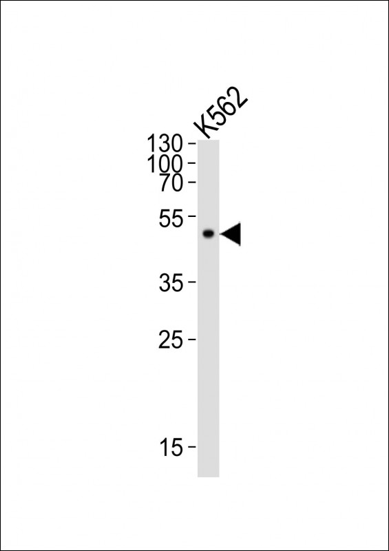
Western blot analysis of lysate from K562 cell line, using ATG4A Antibody(Cat. #P32978). P32978 was diluted at 1:500. A goat anti-mouse IgG H&L(HRP) at 1:10000 dilution was used as the secondary antibody. Lysate at 20μg.
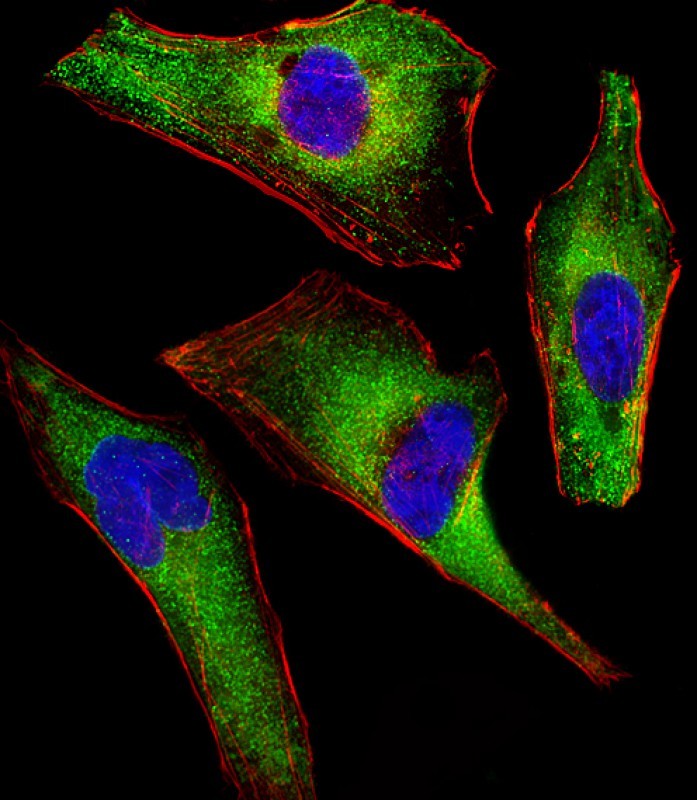
Immunofluorescent analysis of 4% paraformaldehyde-fixed, 0.1% Triton X-100 permeabilized HeLa (human cervical epithelial adenocarcinoma cell line) cells labeling ATG4A with P32978 at 1/25 dilution, followed by Dylight® 488-conjugated goat anti-mouse IgG secondary antibody at 1/200 dilution (green). Immunofluorescence image showing cytoplasm staining on HeLa cell line. Cytoplasmic actin is detected with Dylight® 554 Phalloidin at 1/100 dilution (red).The nuclear counter stain is DAPI (blue).
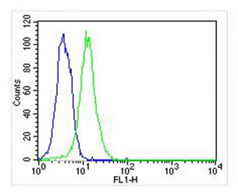
Overlay histogram showing Hela cells stained with P32978 (green line). The cells were fixed with 2% paraformaldehyde (10 min) and then permeabilized with 90% methanol for 10 min. The cells were then icubated in 2% bovine serum albumin to block non-specific protein-protein interactions followed by the antibody (P32978, 1:25 dilution) for 60 min at 37ºC. The secondary antibody used was Goat-Anti-Mouse IgG, DyLight® 488 Conjugated Highly Cross-Adsorbed(NA168821)) at 1/400 dilution for 40 min at 37ºC. Isotype control antibody (blue line) was mouse IgG2b (1μg/1x10^6 cells) used under the same conditions. Acquisition of >10, 000 events was performed.
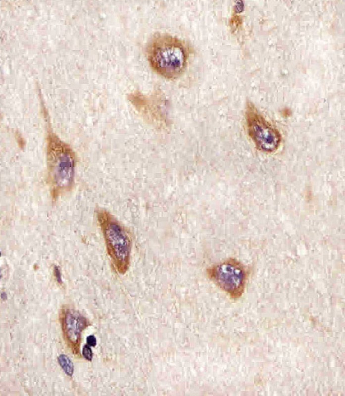
P32978 staining ATG4A in human brain sections by Immunohistochemistry (IHC-P - paraformaldehyde-fixed, paraffin-embedded sections). Tissue was fixed with formaldehyde and blocked with 3% BSA for 0. 5 hour at room temperature; antigen retrieval was by heat mediation with a citrate buffer (pH6). Samples were incubated with primary antibody (1/25) for 1 hours at 37°C. A undiluted biotinylated goat polyvalent antibody was used as the secondary antibody.
Mouse Monoclonal Antibody to ATG4A
-
货号:
P32978 -
别名:
Cysteine protease ATG4A, 3422-, AUT-like 2 cysteine endopeptidase, Autophagin-2, Autophagy-related cysteine endopeptidase 2, Autophagy-related protein 4 homolog A, hAPG4A, ATG4A, APG4A, AUTL2 -
应用:
WB,IHC-P,IF,FCM -
反应种属:
Human -
抗体类型:
Primary antibody -
Swissprot:
Q8WYN0 -
规格:
-
数量:
-+ -
说明书:

目录价¥2180


Mouse Monoclonal Antibody to ATG4A
Description |
|---|
Cysteine protease required for the cytoplasm to vacuole transport (Cvt) and autophagy. Cleaves the C-terminal amino acid of ATG8 family proteins to reveal a C-terminal glycine. Exposure of the glycine at the C-terminus is essential for ATG8 proteins conjugation to phosphatidylethanolamine (PE) and insertion to membranes, which is necessary for autophagy. Preferred substrate is GABARAPL2 followed by MAP1LC3A and GABARAP. Has also an activity of delipidating enzyme for the PE-conjugated forms. |
Specification |
|
|---|---|
| Aliases | Cysteine protease ATG4A, 3422-, AUT-like 2 cysteine endopeptidase, Autophagin-2, Autophagy-related cysteine endopeptidase 2, Autophagy-related protein 4 homolog A, hAPG4A, ATG4A, APG4A, AUTL2 |
| Entrez GeneID | 115201 |
| Swissprot | Q8WYN0 |
| WB Predicted band size | 45.3kDa |
| Host/Isotype | Mouse IgG2b |
| Antibody Type | Primary antibody |
| Storage | Store at 4°C short term. Aliquot and store at -20°C long term. Avoid freeze/thaw cycles. |
| Species Reactivity | Human |
| Immunogen | This ATG4A antibody is generated from a mouse immunized with a recombinant protein. |
Application |
|
|---|---|
| WB | 1/500 |
| IHC | 1/100-1/500 |
| IF/ICC | 1/25 |
| FCM | 1/25 |
Product Image
- Western blot analysis of lysate from K562 cell line, using ATG4A Antibody(Cat. #P32978). P32978 was diluted at 1:500. A goat anti-mouse IgG H&L(HRP) at 1:10000 dilution was used as the secondary antibody. Lysate at 20μg.
- Immunofluorescent analysis of 4% paraformaldehyde-fixed, 0.1% Triton X-100 permeabilized HeLa (human cervical epithelial adenocarcinoma cell line) cells labeling ATG4A with P32978 at 1/25 dilution, followed by Dylight® 488-conjugated goat anti-mouse IgG secondary antibody at 1/200 dilution (green). Immunofluorescence image showing cytoplasm staining on HeLa cell line. Cytoplasmic actin is detected with Dylight® 554 Phalloidin at 1/100 dilution (red).The nuclear counter stain is DAPI (blue).
- Overlay histogram showing Hela cells stained with P32978 (green line). The cells were fixed with 2% paraformaldehyde (10 min) and then permeabilized with 90% methanol for 10 min. The cells were then icubated in 2% bovine serum albumin to block non-specific protein-protein interactions followed by the antibody (P32978, 1:25 dilution) for 60 min at 37ºC. The secondary antibody used was Goat-Anti-Mouse IgG, DyLight® 488 Conjugated Highly Cross-Adsorbed(NA168821)) at 1/400 dilution for 40 min at 37ºC. Isotype control antibody (blue line) was mouse IgG2b (1μg/1x10^6 cells) used under the same conditions. Acquisition of >10, 000 events was performed.
- P32978 staining ATG4A in human brain sections by Immunohistochemistry (IHC-P - paraformaldehyde-fixed, paraffin-embedded sections). Tissue was fixed with formaldehyde and blocked with 3% BSA for 0. 5 hour at room temperature; antigen retrieval was by heat mediation with a citrate buffer (pH6). Samples were incubated with primary antibody (1/25) for 1 hours at 37°C. A undiluted biotinylated goat polyvalent antibody was used as the secondary antibody.
For Reseach Only
Application Key:WB - Western Blot | IHC - Immunohistochemistry | ICC - Immunocytochemistry | FCM - Flow Cytometry | ELISA - Enzyme-linked Immunosorbent Assay | IP - Immunoprecipitation
#P32978

相关产品


联系方式
CONTACT-
联系电话:
0731-88388785 -
公司邮箱:
sales@promab.cn -
公司地址:
湖南省长沙市高新开发区林语路239号顺畅产业园5楼
 官方微信
官方微信
产品中心
Product技术服务
service客户留言
message