


提醒成功


 微信/QQ登录
搜索


 首页
首页
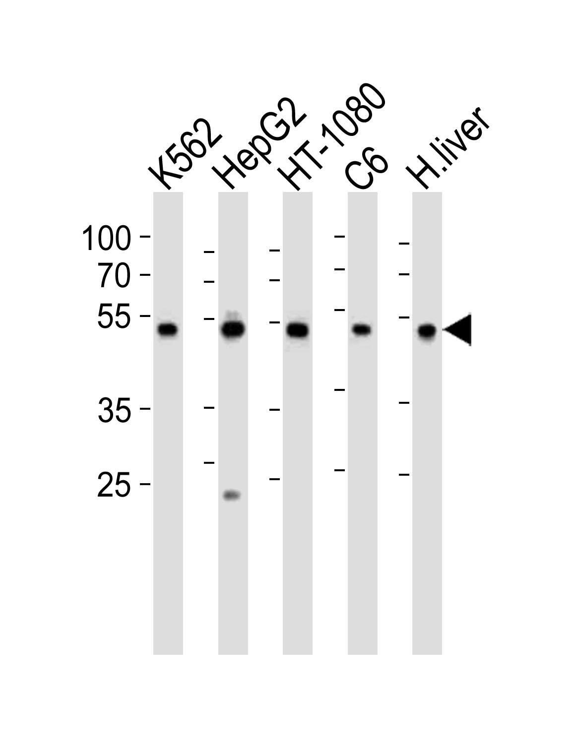
Western blot analysis of lysates from K562, HepG2, HT-1080, rat C6 cell line and human liver tissue lysate (from left to right), using PDIA6 Antibody (Center K159)(Cat. #P32745). P32745 was diluted at 1:1000 at each lane. A goat anti-rabbit IgG H&L(HRP) at 1:10000 dilution was used as the secondary antibody. Lysates at 35ug per lane.
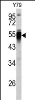
Western blot analysis of PDIA6 antibody (Center K159) (Cat.# P32745) in Y79 cell line lysates (35ug/lane). PDIA6 (arrow) was detected using the purified Pab.
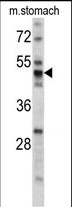
Western blot analysis of PDIA6 antibody (Center K159) (Cat.# P32745) in mouse stomach tissue lysates (35ug/lane). PDIA6 (arrow) was detected using the purified Pab.
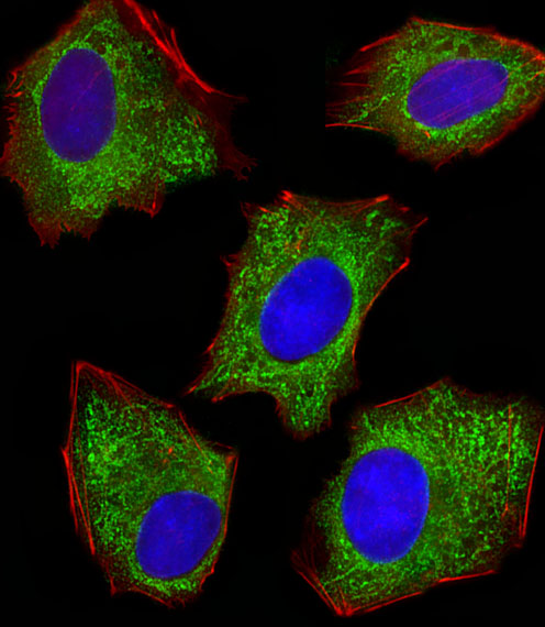
Fluorescent image of HepG2 cells stained with XAF1 PDIA6 Antibody (Center K159)(Cat#P32745). P32745 was diluted at 1:100 dilution. An Alexa Fluor 488-conjugated goat anti-rabbit lgG at 1:400 dilution was used as the secondary antibody (green). DAPI was used to stain the cell nuclear (blue). Cytoplasmic actin was counterstained with Alexa Fluor® 555 conjugated with Phalloidin (red).
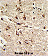
Formalin-fixed and paraffin-embedded human brain tissue reacted with PDIA6 Antibody (Center K159), which was peroxidase-conjugated to the secondary antibody, followed by DAB staining. This data demonstrates the use of this antibody for immunohistochemistry; clinical relevance has not been evaluated.
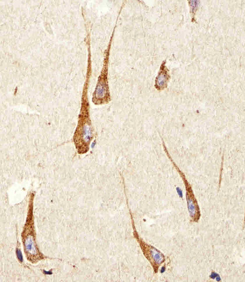
Immunohistochemical analysis of paraffin-embedded H. brain section using PDIA6 Antibody (Center K159)(Cat#P32745). P32745 was diluted at 1:100 dilution. A peroxidase-conjugated goat anti-rabbit IgG at 1:400 dilution was used as the secondary antibody, followed by DAB staining.
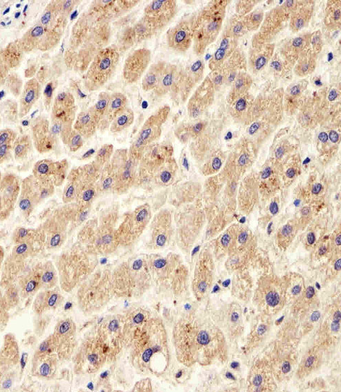
Immunohistochemical analysis of paraffin-embedded H. liver section using PDIA6 Antibody (Center K159)(Cat#P32745). P32745 was diluted at 1:100 dilution. A peroxidase-conjugated goat anti-rabbit IgG at 1:400 dilution was used as the secondary antibody, followed by DAB staining.
Rabbit Polyclonal Antibody to PDIA6 (Center K159)
-
货号:
P32745 -
别名:
Protein disulfide-isomerase A6, Endoplasmic reticulum protein 5, ER protein 5, ERp5, Protein disulfide isomerase P5, Thioredoxin domain-containing protein 7, PDIA6, ERP5, P5, TXNDC7 -
应用:
WB,IHC-P,IF -
反应种属:
Human, Mouse, Rat -
抗体类型:
Primary antibody -
Swissprot:
Q15084 -
规格:
-
数量:
-+ -
说明书:

目录价¥1980


Rabbit Polyclonal Antibody to PDIA6 (Center K159)
Description |
|---|
Protein disulfide isomerases (EC 5.3.4.1), such as PDIA6, are endoplasmic reticulum (ER) resident proteins that catalyze formation, reduction, and isomerization of disulfide bonds in proteins and are thought to play a role in folding of disulfide-bonded proteins. |
Specification |
|
|---|---|
| Aliases | Protein disulfide-isomerase A6, Endoplasmic reticulum protein 5, ER protein 5, ERp5, Protein disulfide isomerase P5, Thioredoxin domain-containing protein 7, PDIA6, ERP5, P5, TXNDC7 |
| Entrez GeneID | 10130 |
| Swissprot | Q15084 |
| WB Predicted band size | 48.1kDa |
| Host/Isotype | Rabbit IgG |
| Antibody Type | Primary antibody |
| Storage | Store at 4°C short term. Aliquot and store at -20°C long term. Avoid freeze/thaw cycles. |
| Species Reactivity | Human, Mouse, Rat |
| Immunogen | This PDIA6 antibody is generated from rabbits immunized with a KLH conjugated synthetic peptide between 144-172 amino acids from the Central region of human PDIA6. |
| Formulation | Purified antibody in PBS with 0.05% sodium azide,1%BSA and 50% glycerol.prepared by Saturated Ammonium Sulfate (SAS) . |
Application |
|
|---|---|
| WB | 1/1000 |
| IHC | 1/100-1/500 |
| IF/ICC | 1/100 |
Product Image
- Western blot analysis of lysates from K562, HepG2, HT-1080, rat C6 cell line and human liver tissue lysate (from left to right), using PDIA6 Antibody (Center K159)(Cat. #P32745). P32745 was diluted at 1:1000 at each lane. A goat anti-rabbit IgG H&L(HRP) at 1:10000 dilution was used as the secondary antibody. Lysates at 35ug per lane.
- Western blot analysis of PDIA6 antibody (Center K159) (Cat.# P32745) in Y79 cell line lysates (35ug/lane). PDIA6 (arrow) was detected using the purified Pab.
- Western blot analysis of PDIA6 antibody (Center K159) (Cat.# P32745) in mouse stomach tissue lysates (35ug/lane). PDIA6 (arrow) was detected using the purified Pab.
- Fluorescent image of HepG2 cells stained with XAF1 PDIA6 Antibody (Center K159)(Cat#P32745). P32745 was diluted at 1:100 dilution. An Alexa Fluor 488-conjugated goat anti-rabbit lgG at 1:400 dilution was used as the secondary antibody (green). DAPI was used to stain the cell nuclear (blue). Cytoplasmic actin was counterstained with Alexa Fluor® 555 conjugated with Phalloidin (red).
- Formalin-fixed and paraffin-embedded human brain tissue reacted with PDIA6 Antibody (Center K159), which was peroxidase-conjugated to the secondary antibody, followed by DAB staining. This data demonstrates the use of this antibody for immunohistochemistry; clinical relevance has not been evaluated.
- Immunohistochemical analysis of paraffin-embedded H. brain section using PDIA6 Antibody (Center K159)(Cat#P32745). P32745 was diluted at 1:100 dilution. A peroxidase-conjugated goat anti-rabbit IgG at 1:400 dilution was used as the secondary antibody, followed by DAB staining.
- Immunohistochemical analysis of paraffin-embedded H. liver section using PDIA6 Antibody (Center K159)(Cat#P32745). P32745 was diluted at 1:100 dilution. A peroxidase-conjugated goat anti-rabbit IgG at 1:400 dilution was used as the secondary antibody, followed by DAB staining.
For Reseach Only
Application Key:WB - Western Blot | IHC - Immunohistochemistry | ICC - Immunocytochemistry | FCM - Flow Cytometry | ELISA - Enzyme-linked Immunosorbent Assay | IP - Immunoprecipitation
#P32745

相关产品


联系方式
CONTACT-
联系电话:
0731-88388785 -
公司邮箱:
sales@promab.cn -
公司地址:
湖南省长沙市高新开发区林语路239号顺畅产业园5楼
 官方微信
官方微信
产品中心
Product技术服务
service客户留言
message