


提醒成功


 微信/QQ登录
搜索


 首页
首页
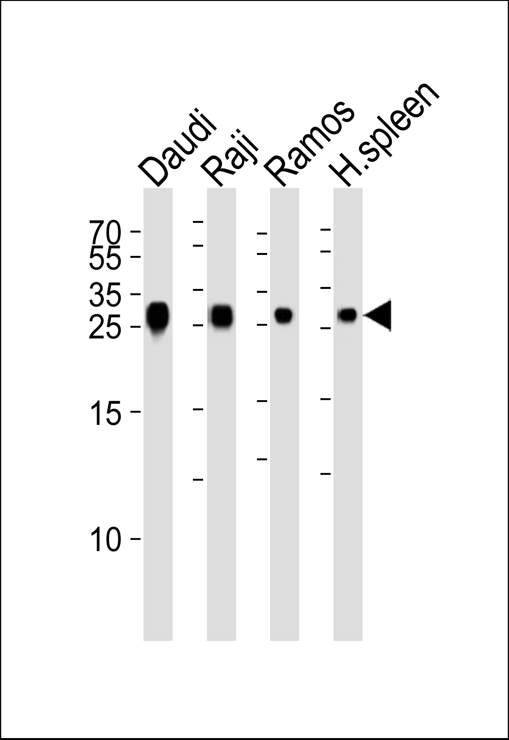
Western blot analysis of lysates from Daudi, Raji, Ramos cell line and houman spleen tissue lysate (from left to right), using HLA-DRB1 Antibody (Center) (Cat. #P32609). P32609 was diluted at 1:1000 at each lane. A goat anti-rabbit IgG H&L(HRP) at 1:5000 dilution was used as the secondary antibody. Lysates at 35ug per lane.
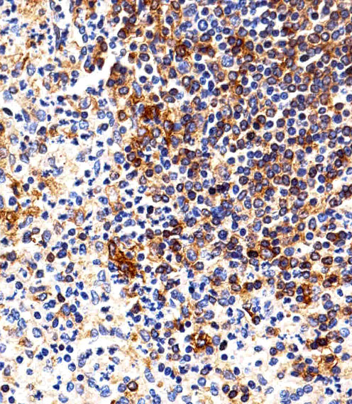
Immunohistochemical analysis of paraffin-embedded H. spleen section using HLA-DRB1 Antibody (Center)(Cat#P32609). P32609 was diluted at 1:25 dilution. A peroxidase-conjugated goat anti-rabbit IgG at 1:400 dilution was used as the secondary antibody, followed by DAB staining.

Immunohistochemical analysis of paraffin-embedded H. tonsil section using HLA-DRB1 Antibody (Center)(Cat#P32609). P32609 was diluted at 1:25 dilution. A peroxidase-conjugated goat anti-rabbit IgG at 1:400 dilution was used as the secondary antibody, followed by DAB staining.
Rabbit Polyclonal Antibody to HLA-DRB1
-
货号:
P32609 -
别名:
HLA class II histocompatibility antigen, DRB1-1 beta chain, MHC class II antigen DRB1*1, DR-1, DR1, HLA-DRB1 -
应用:
WB,IHC-P -
反应种属:
Human -
抗体类型:
Primary antibody -
Swissprot:
P04229 -
规格:
-
数量:
-+ -
说明书:

目录价¥1980


Rabbit Polyclonal Antibody to HLA-DRB1
Description |
|---|
Binds peptides derived from antigens that access the endocytic route of antigen presenting cells (APC) and presents them on the cell surface for recognition by the CD4 T-cells. The peptide binding cleft accommodates peptides of 10-30 residues. The peptides presented by MHC class II molecules are generated mostly by degradation of proteins that access the endocytic route; where they are processed by lysosomal proteases and other hydrolases. Exogenous antigens that have been endocytosed by the APC are thus readily available for presentation via MHC II molecules; and for this reason this antigen presentation pathway is usually referred to as exogenous. As membrane proteins on their way to degradation in lysosomes as part of their normal turn-over are also contained in the endosomal/lysosomal compartments; exogenous antigens must compete with those derived from endogenous components. Autophagy is also a source of endogenous peptides; autophagosomes constitutively fuse with MHC class II loading compartments. In addition to APCs; other cells of the gastrointestinal tract; such as epithelial cells; express MHC class II molecules and CD74 and act as APCs; which is an unusual trait of the GI tract. To produce a MHC class II molecule that presents an antigen; three MHC class II molecules (heterodimers of an alpha and a beta chain) associate with a CD74 trimer in the ER to form a heterononamer. Soon after the entry of this complex into the endosomal/lysosomal system where antigen processing occurs; CD74 undergoes a sequential degradation by various proteases; including CTSS and CTSL; leaving a small fragment termed CLIP (class-II-associated invariant chain peptide). The removal of CLIP is facilitated by HLA-DM via direct binding to the alpha-beta-CLIP complex so that CLIP is released. HLA-DM stabilizes MHC class II molecules until primary high affinity antigenic peptides are bound. The MHC II molecule bound to a peptide is then transported to the cell membrane surface. In B-cells; the interaction between HLA-DM and MHC class II molecules is regulated by HLA-DO. Primary dendritic cells (DCs) also to express HLA-DO. Lysosomal microenvironment has been implicated in the regulation of antigen loading into MHC II molecules; increased acidification produces increased proteolysis and efficient peptide loading. |
Specification |
|
|---|---|
| Aliases | HLA class II histocompatibility antigen, DRB1-1 beta chain, MHC class II antigen DRB1*1, DR-1, DR1, HLA-DRB1 |
| Swissprot | P04229 |
| Host/Isotype | Rabbit IgG |
| Antibody Type | Primary antibody |
| Storage | Store at 4°C short term. Aliquot and store at -20°C long term. Avoid freeze/thaw cycles. |
| Species Reactivity | Human |
| Immunogen | This HLA-DRB1 antibody is generated from a rabbit immunized with a KLH conjugated synthetic peptide between 103-137 amino acids from the Central region of human HLA-DRB1. |
Application |
|
|---|---|
| WB | 1/1000 |
| IHC | 1/100-1/500 |
Product Image
- Western blot analysis of lysates from Daudi, Raji, Ramos cell line and houman spleen tissue lysate (from left to right), using HLA-DRB1 Antibody (Center) (Cat. #P32609). P32609 was diluted at 1:1000 at each lane. A goat anti-rabbit IgG H&L(HRP) at 1:5000 dilution was used as the secondary antibody. Lysates at 35ug per lane.
- Immunohistochemical analysis of paraffin-embedded H. spleen section using HLA-DRB1 Antibody (Center)(Cat#P32609). P32609 was diluted at 1:25 dilution. A peroxidase-conjugated goat anti-rabbit IgG at 1:400 dilution was used as the secondary antibody, followed by DAB staining.
- Immunohistochemical analysis of paraffin-embedded H. tonsil section using HLA-DRB1 Antibody (Center)(Cat#P32609). P32609 was diluted at 1:25 dilution. A peroxidase-conjugated goat anti-rabbit IgG at 1:400 dilution was used as the secondary antibody, followed by DAB staining.
For Reseach Only
Application Key:WB - Western Blot | IHC - Immunohistochemistry | ICC - Immunocytochemistry | FCM - Flow Cytometry | ELISA - Enzyme-linked Immunosorbent Assay | IP - Immunoprecipitation
#P32609

相关产品


联系方式
CONTACT-
联系电话:
0731-88388785 -
公司邮箱:
sales@promab.cn -
公司地址:
湖南省长沙市高新开发区林语路239号顺畅产业园5楼
 官方微信
官方微信
产品中心
Product技术服务
service客户留言
message